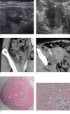
Signet-ring cell carcinoma of the appendix: a case report with an emphasis on sonographic findings
- PMID: 26753605
- PMCID: PMC4825206
- DOI: 10.14366/usg.15063
Signet-ring cell carcinoma of the appendix: a case report with an emphasis on sonographic findings
Abstract
In this report, we present a rare case of primary signet-ring cell carcinoma of the appendix in a 51-year-old woman with right lower quadrant pain. Since non-specific concentric appendiceal wall thickening was found in a radiologic evaluation, it was misdiagnosed as non-tumorous appendicitis. An in-depth examination of the correlation between sonographic and histopathologic findings demonstrated that a single markedly thickened hypoechoic layer was well correlated with the diffuse infiltration of tumor cells in both the submucosal and muscle layers. If this sonographic finding is observed in certain clinical settings, such as potential ovarian and peritoneal metastasis, submucosal infiltrative tumors, including signet-ring cell carcinoma, should be considered in the differential diagnosis.
Keywords: Appendix; Carcinoma, signet ring cell; Ultrasonography.
Conflict of interest statement
No potential conflict of interest relevant to this article was reported.
Figures

Similar articles
-
Appendiceal Signet Ring Cell Carcinoma Presenting As Acute Appendicitis: A Case Report.Cureus. 2024 Apr 27;16(4):e59137. doi: 10.7759/cureus.59137. eCollection 2024 Apr. Cureus. 2024. PMID: 38803764 Free PMC article.
-
Signet Ring Carcinoma of the Appendix Presenting as Crohn's Disease in a Young Male.Case Rep Gastroenterol. 2018 Jun 15;12(2):277-285. doi: 10.1159/000489298. eCollection 2018 May-Aug. Case Rep Gastroenterol. 2018. PMID: 30022916 Free PMC article.
-
Primary signet ring cell carcinoma of the appendix: A rare case report.World J Clin Cases. 2015 Jun 16;3(6):538-41. doi: 10.12998/wjcc.v3.i6.538. World J Clin Cases. 2015. PMID: 26090376 Free PMC article.
-
[Experience of the Pharmacotherapy against Appendix and Sigmoid Colon Signet Ring Cell Carcinoma with the Peritoneal Dissemination].Gan To Kagaku Ryoho. 2015 Oct;42(10):1268-70. Gan To Kagaku Ryoho. 2015. PMID: 26489568 Review. Japanese.
-
Primary appendiceal adenocarcinoma.Am J Clin Oncol. 1999 Oct;22(5):458-9. doi: 10.1097/00000421-199910000-00007. Am J Clin Oncol. 1999. PMID: 10521058 Review.
Cited by
-
Clinical benefits of FOLFOXIRI combined with bevacizumab for advanced-stage primary signet ring cell carcinoma of the appendix: A case report.Medicine (Baltimore). 2023 Aug 4;102(31):e34412. doi: 10.1097/MD.0000000000034412. Medicine (Baltimore). 2023. PMID: 37543827 Free PMC article.
-
Appendiceal Signet Ring Cell Carcinoma Presenting As Acute Appendicitis: A Case Report.Cureus. 2024 Apr 27;16(4):e59137. doi: 10.7759/cureus.59137. eCollection 2024 Apr. Cureus. 2024. PMID: 38803764 Free PMC article.
-
Sonographic Findings of Malignant Appendix Tumors in Seven Cases.J Med Ultrasound. 2018 Jan-Mar;26(1):52-55. doi: 10.4103/JMU.JMU_16_17. Epub 2018 Mar 28. J Med Ultrasound. 2018. PMID: 30065515 Free PMC article.
-
Appendiceal Signet Ring Cell Carcinoma: An Atypical Cause of Acute Appendicitis-A Case Study and Review of Current Knowledge.Diagnostics (Basel). 2023 Jul 13;13(14):2359. doi: 10.3390/diagnostics13142359. Diagnostics (Basel). 2023. PMID: 37510102 Free PMC article.
References
-
- Pickhardt PJ, Levy AD, Rohrmann CA, Jr, Kende AI. Primary neoplasms of the appendix manifesting as acute appendicitis: CT findings with pathologic comparison. Radiology. 2002;224:775–781. - PubMed
-
- Pickhardt PJ, Levy AD, Rohrmann CA, Jr, Kende AI. Primary neoplasms of the appendix: radiologic spectrum of disease with pathologic correlation. Radiographics. 2003;23:645–662. - PubMed
Publication types
LinkOut - more resources
Full Text Sources
Other Literature Sources

