. 2016 Jan 12:6:19020.
doi: 10.1038/srep19020.

Characterization of the interactions of PARP-1 with UV-damaged DNA in vivo and in vitro

Affiliations

Characterization of the interactions of PARP-1 with UV-damaged DNA in vivo and in vitro

Nupur K Purohit et al. Sci Rep. .

Abstract

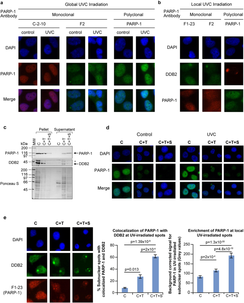
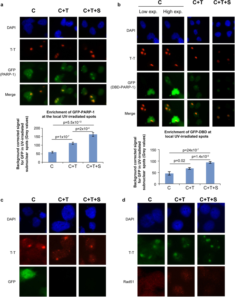
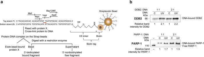
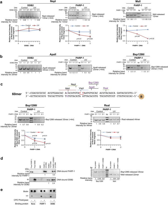
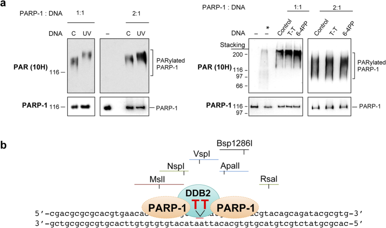
The existing methodologies for studying robust responses of poly (ADP-ribose) polymerase-1 (PARP-1) to DNA damage with strand breaks are often not suitable for examining its subtle responses to altered DNA without strand breaks, such as UV-damaged DNA. Here we describe two novel assays with which we characterized the interaction of PARP-1 with UV-damaged DNA in vivo and in vitro. Using an in situ fractionation technique to selectively remove free PARP-1 while retaining the DNA-bound PARP-1, we demonstrate a direct recruitment of the endogenous or exogenous PARP-1 to the UV-lesion site in vivo after local irradiation. In addition, using the model oligonucleotides with single UV lesion surrounded by multiple restriction enzyme sites, we demonstrate in vitro that DDB2 and PARP-1 can simultaneously bind to UV-damaged DNA and that PARP-1 casts a bilateral asymmetric footprint from -12 to +9 nucleotides on either side of the UV-lesion. These techniques will permit characterization of different roles of PARP-1 in the repair of UV-damaged DNA and also allow the study of normal housekeeping roles of PARP-1 with undamaged DNA.


Figures

Figure 1
Figure 1. In situ fractionation to reveal the recruitment of endogenous PARP-1 to UV-induced DNA lesion site.
(a,b) Unchanged pattern of nuclear staining for PARP-1 after global or local UVC-irradiation of cells processed with conventional immunocytological techniques. Human skin fibroblasts were exposed either to global (panel a) or local (panel b) irradiation with UVC, fixed with formaldehyde-methanol and probed for PARP-1 (global and local UVC) and DDB2 (local UVC) using specified antibodies. DAPI staining was carried out to define nuclei. (c) Efficiency of extraction of free PARP-1 and DDB2 from adherent control GMU6 cells. The pellets and supernatants obtained from equivalent cell numbers after extraction with CSK buffer (C), CSK+0.5% Triton (C+T) or CSK + 0.5% Triton + 0.42 M NaCl (C+T+S) were immunoblotted for PARP-1 and DDB2. The *refers to non-specific signal in DDB2 probing and Ponceau S staining reflected the residual protein content in cell pellets and supernatant at the end of each protocol. (d) Comparison of the efficiency of three protocols for extraction of the endogenous PARP-1 from adherent control and UV-irradiated cells. The GMU6 human skin fibroblasts were globally irradiated with 10 J/m2 UVC (or control), extracted 10 min later with the three protocols and probed for PARP-1 using polyclonal PARP-1 antibody. (e) Colocalization of endogenous PARP-1 with DDB2 at local UVC-induced DNA damage. GMU6 cells were irradiated locally with 100 J/m2 and after 10 min subjected to the three protocols (C, C+T and C+T+S) followed by visualization of PARP-1 (F1-23, red) and DDB2 (green). The left chart represents the percent of subnuclear PARP-1 spots that colocalize with DDB2. The right chart represents the quantification of the PARP-1 intensity at the DDB2 spots after subtraction of background signal intensity for PARP-1 from an equivalent area of unirradiated part of the same nucleus. Data of the charts are pooled from three experiments (n = 120–150 spots, data points are mean ± s.e.).
Figure 2
Figure 2. In situ fractionation improves detection of exogenous GFP-PARP-1 or its DNA binding domain at local UV-irradiated spots.
(a,b) Recruitment of GFP-PARP-1 or its DBD to UV-induced T-T lesions. GMU6 cells were transfected with GFP- PARP-1 or GFP-DBD of PARP-1 for 24 h. The cells were locally irradiated and processed by C, C+T or C+T+S protocols. GFP-PARP-1 or GFP-DBD (green) and T-T (red) were visualized in DAPI-stained nuclei by immunofluorescence microscopy. The charts represent the quantification of GFP intensity for GFP- PARP-1 or GFP-DBD of PARP-1 at the T-T spots after background correction. (n = 80–150 spots, data points are mean ± s.e.). (c,d) Specificity of in situ extraction protocol: unrelated nuclear proteins (GFP and Rad51) do not colocalize with UV-damaged DNA. GMU6 cells were locally irradiated with 100 J/m2 UVC and subjected 10 min later to in situ fractionation using the three protocols. For panel-c, the cells were transfected with GFP expressing plasmid 24 h before irradiation and protein extraction. The cells were processed for immunofluorescence detection of GFP, Rad51 (green) and T-T (red). DAPI staining was carried out to define nuclei.
Figure 3
Figure 3. Strategy to study binding and footprint of proteins on UV-DNA.
(a) The experimental design for determining the extent of binding of proteins to UV-DNA and analyses of protection of DNA during restriction digestion. The control and UV-DNA were immobilized on streptavidin beads via their biotin tag and reacted with purified PARP-1 or DDB2. The unreacted proteins were removed and bound proteins were cross-linked. The beads were then either analysed for bound-proteins by eluting the protein in Laemmli buffer, followed by SDS-PAGE, transfer and probe with specific antibodies or they were subjected to restriction digestion followed by analyses of the released DNA fragments on 10–15% native PAGE stained with gel red. (b) DDB2 and PARP-1 bind more to UV-DNA than control DNA. PARP-1 and DDB2 were reacted with 50 ng of control or UV-DNA at 1:1 or 1:2 (DNA:protein) molar ratios. The proteins were eluted from the beads, resolved on SDS-PAGE and analyzed by immunoblotting as shown in (a). The band intensities of protein bound to UV-DNA are shown as relative to signal for protein bound to control DNA.
Figure 4
Figure 4. Footprinting of PARP-1 and DDB2 at UV-lesion site.
(a) Restriction mapping of proteins on the 5′of the UV-lesion on 40mer DNA. 100 ng of bead-bound control or UV-DNA were reacted with DDB2 or PARP-1 at different DNA: protein ratios and digested at 37 °C with NspI (30 min) or MslI (15 min). The released 5′-fragments were resolved on 15% native PAGE and band intensities were measured. Each data point derived from three independent experiments represents mean ± s.d. for relative band intensity from three experiments for the fragment released from protein-bound versus protein-free DNA, with P values shown in the chart. (b) Mapping of proteins on the 3′-side of the UV-lesion on 40mer DNA. The protein-bound DNA was digested with ApalI (60 min) and Bsp1286I (20 min), and released 5′-fragments were resolved on 12% native PAGE. The data derived from two independent experiments is represented in the chart as described in panel-a. (c) Top panel-Structure of 60mer oligo with defined UV-damage. The 60mer oligo sequence was based on 40mer oligo but with a new RsaI site near 3′-end. Bottom panels- Restriction mapping of proteins on the 3′-side of the UV-lesion on 60mer DNA. The protein-bound DNA was digested with Bsp1286I (20 min) and RsaI (30 min) and released 5′-fragments were analysed by 12% native PAGE. The data derived from three independent experiments is represented in the chart as described in panel-a. (d) Simultaneous binding and footprint of DDB2 and PARP-1 on 60mer UV-DNA. PARP-1 and DDB2 were reacted either separately or together with bead-bound UV-DNA (50 ng), at 2:1 molar ratio of protein:DNA. The proteins bound to the beads were detected by immunoblotting (left panels), and footprint of proteins on DNA was examined by restriction digestion with Bsp1286I and Msll (right panels). (e) Repair of T-T by CPD photolyase despite binding of DDB2 or PARP-1 to UV-DNA. Bead-bound 40mer UV-DNA was reacted (or not) with DDB2 or PARP-1, and subjected to repair (or not) by CPD photolyase. The DNA was eluted and immunodot-blotted for T-T. The data represents identical results obtained in three independent experiments.
Figure 5
Figure 5. Catalytic activation of PARP-1 with defined UV-damaged DNA and a model for footprint of PARP-1 and DDB2 on UV-lesion site.
(a) Stimulation of catalytic activity of PARP-1 by various defined UV-damaged DNA in vitro. PARP-1 activation assay was performed using 40 mer control or UV-DNA (left panel) or using 24mer DNA with no damage (control) or with a single defined T-T and 6–4PP (right panel) at 1:1 or 1:2 molar ratio of DNA:protein. After resolution on SDS-PAGE, immunoprobing for PARP-1 and PARylated PARP-1 (10H antibody) was carried out. The *refers to the cell extract containing H2O2-activated PARP-1. Panel represents one of three identical experiments. (b) Model for binding of PARP-1 and DDB2 to the UV-lesion site on 60mer oligo (see Results and Discussion section for details).

References

    1. Robert I., Karicheva O., Reina San Martin B., Schreiber V. & Dantzer F. Functional aspects of PARylation in induced and programmed DNA repair processes: Preserving genome integrity and modulating physiological events. Mol. Aspects Med. 34, 1138–1152 (2013). - PubMed
    1. Pascal J. M. & Ellenberger T. The rise and fall of poly(ADP-ribose): An enzymatic perspective. DNA Repair (Amst) 32, 10–16 (2015). - PMC - PubMed
    1. Kim M. Y., Mauro S., Gevry N., Lis J. T. & Kraus W. L. NAD+-dependent modulation of chromatin structure and transcription by nucleosome binding properties of PARP-1. Cell 119, 803–814 (2004). - PubMed
    1. Ghodgaonkar M. M., Zacal N. J., Kassam S. N., Rainbow A. J. & Shah G. M. Depletion of poly(ADP-ribose) polymerase-1 reduces host cell reactivation for UV-treated adenovirus in human dermal fibroblasts. DNA Repair (Amst) 7, 617–632 (2008). - PubMed
    1. Pines A. et al. PARP1 promotes nucleotide excision repair through DDB2 stabilization and recruitment of ALC1. J. Cell Biol. 199, 235–249 (2012). - PMC - PubMed

Publication types

MeSH terms

LinkOut - more resources