Review
. 2016 Mar;53(3):145-51.
doi: 10.1136/jmedgenet-2015-103387. Epub 2016 Jan 11.

The importance of genetic diagnosis for Duchenne muscular dystrophy

Affiliations
Review

The importance of genetic diagnosis for Duchenne muscular dystrophy

Annemieke Aartsma-Rus et al. J Med Genet. 2016 Mar.

Abstract

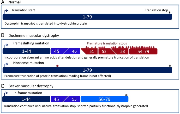
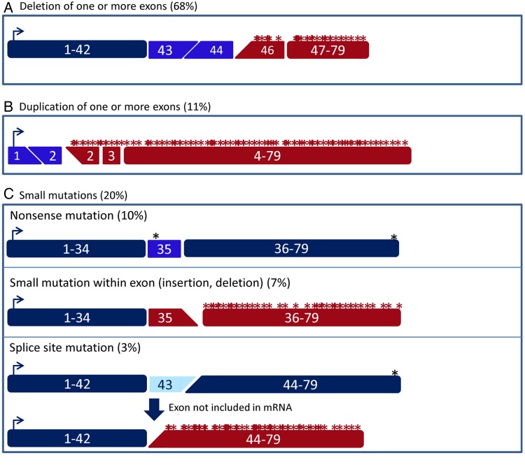
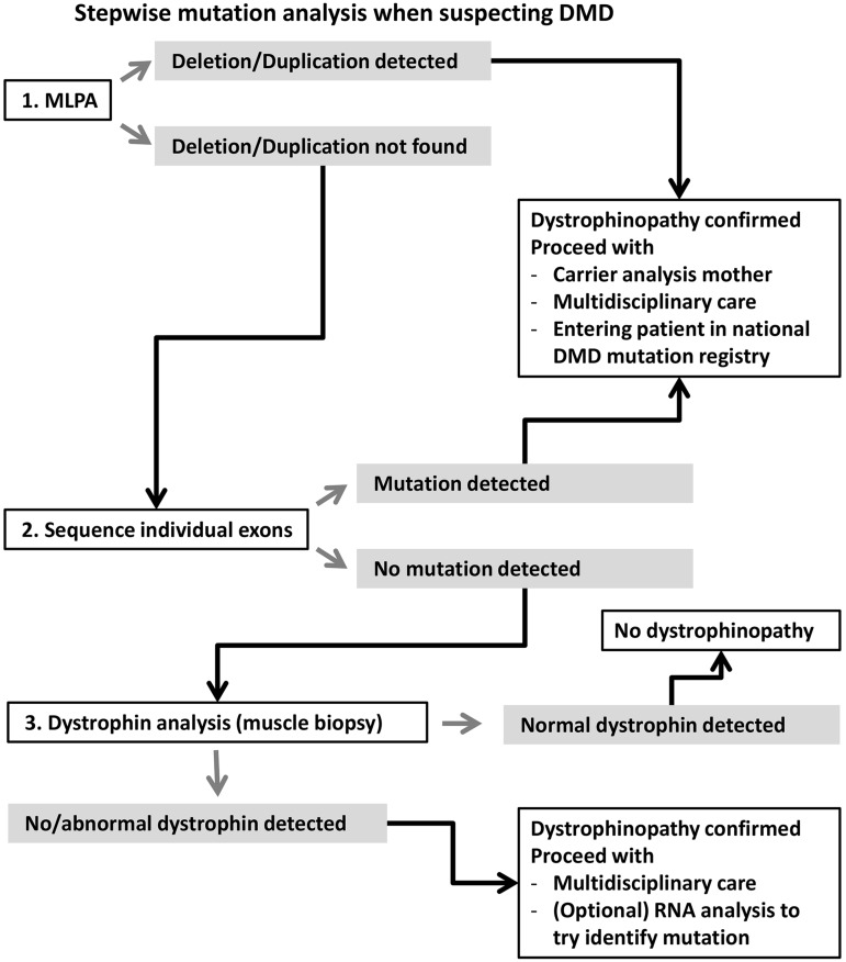
Duchenne muscular dystrophy (DMD) and Becker muscular dystrophy are caused by mutations in the dystrophin-encoding DMD gene. Large deletions and duplications are most common, but small mutations have been found as well. Having a correct diagnosis is important for family planning and providing proper care to patients according to published guidelines. With mutation-specific therapies under development for DMD, a correct diagnosis is now also important for assessing whether patients are eligible for treatments. This review discusses different mutations causing DMD, diagnostic techniques available for making a genetic diagnosis for children suspected of DMD and the importance of having a specific genetic diagnosis in the context of emerging genetic therapies for DMD.

Keywords: Diagnosis; Genetics; Muscle disease.


Figures

Figure 1
Figure 1
Schematic depiction of dystrophin transcripts in healthy, Duchenne muscular dystrophy (DMD) and Becker muscular dystrophy (BMD) individuals. (A) In the normal situation, the dystrophin mRNA consists of 79 exons that are translated into the dystrophin protein. (B) In patients with DMD, protein translation is stopped prematurely. This can be due to frame-shifting mutations (in this example, a deletion of exons 47–50, top panel) that lead to the inclusion of aberrant amino acids and generally premature truncation of translation. Alternatively, a point mutation can change an amino acid codon into a stop codon (bottom panel, nonsense mutation). This premature stop codon will be used instead of the natural stop codon at the end of the transcript. (C) In patients with BMD, mutations maintain the open reading frame (in this example, a deletion of exons 46–54). As such, protein translation does not stop prematurely but continues until the natural stop codon at the end of the mRNA. However, the generated dystrophin will be shorter because it will lack the amino acids encoded by exons 46–54.
Figure 2
Figure 2
Schematic depiction of the effect of different types of Duchenne muscular dystrophy (DMD)-causing mutations on the dystrophin transcript. (A) Deletions of one or more exons can cause a shift of the open reading frame (in this example, a deletion of exon 45). (B) A duplication of one or more exons can cause a shift of the open reading frame (in this example, a duplication of exon 2). (C) There are several types of small mutations than can cause DMD. Nonsense mutations (top panel) introduce a stop codon prematurely (in this example, the nonsense mutation is located in exon 35). Small insertions or deletions (middle panel) can disrupt the open reading frame (in this example, a 1 bp insertion in exon 35). Finally, mutations affecting the splice sites (bottom panel) generally lead to the exclusion of the affected exon from the mRNA (in this example, exon 43). As such, a single-exon deletion that disrupts the open reading frame is generated on the mRNA level.
Figure 3
Figure 3
Overview of suggested stepwise mutation analysis when suspecting Duchenne muscular dystrophy (DMD). When reporting mutations, it is important to use standard nomenclature as described on the website of the Human Genome Variation Society (http://www.hgvs.org/mutnomen/). Furthermore, it is important to include patients in national patient registries (upon consent), so they can be contacted when mutation-specific therapies are tested in clinical trials or become available on the market. The majority of mutations will be detected by multiplex ligation-dependent probe amplification (MLPA) or exon sequencing on DNA level. If no mutation is found, a muscle biopsy can be taken to study dystrophin protein. When dystrophin is abnormal or absent in a muscle biopsy, while no mutation can be found with MLPA or exon sequencing, one can consider analysing muscle RNA to identify the potential inclusion of a cryptic exon due to an intronic mutation (these will not be identified with MLPA or exon sequencing).

References

    1. Emery AE. The muscular dystrophies. Lancet 2002;359:687–95. 10.1016/S0140-6736(02)07815-7 - DOI - PubMed
    1. Flanigan KM. Duchenne and Becker muscular dystrophies. Neurol Clin 2014;32:671–88, viii 10.1016/j.ncl.2014.05.002 - DOI - PubMed
    1. Koenig M, Monaco AP, Kunkel LM. The complete sequence of dystrophin predicts a rod-shaped cytoskeletal protein. Cell 1988;53:219–28. 10.1016/0092-8674(88)90383-2 - DOI - PubMed
    1. Aartsma-Rus A. Dystrophin analysis in clinical trials. J Neuromuscul Dis 2014;1:41–53. - PubMed
    1. Muntoni F, Torelli S, Ferlini A. Dystrophin and mutations: one gene, several proteins, multiple phenotypes. Lancet Neurol 2003;2:731–40. 10.1016/S1474-4422(03)00585-4 - DOI - PubMed

MeSH terms