. 2016 Jan 12:15:4.
doi: 10.1186/s12944-015-0175-2.

Why high cholesterol levels help hematological malignancies: role of nuclear lipid microdomains

Affiliations

Why high cholesterol levels help hematological malignancies: role of nuclear lipid microdomains

Michela Codini et al. Lipids Health Dis. .

Abstract

Background: Diet and obesity are recognized in the scientific literature as important risk factors for cancer development and progression. Hypercholesterolemia facilitates lymphoma lymphoblastic cell growth and in time turns in hypocholesterolemia that is a sign of tumour progression. The present study examined how and where the cholesterol acts in cancer cells when you reproduce in vitro an in vivo hypercholesterolemia condition.

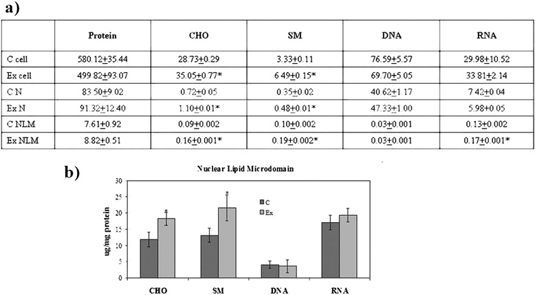
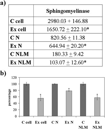
Methods: We used non-Hodgkin's T cell human lymphoblastic lymphoma (SUP-T1 cell line) and we studied cell morphology, aggressiveness, gene expression for antioxidant proteins, polynucleotide kinase/phosphatase and actin, cholesterol and sphingomyelin content and finally sphingomyelinase activity in whole cells, nuclei and nuclear lipid microdomains.

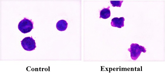
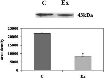
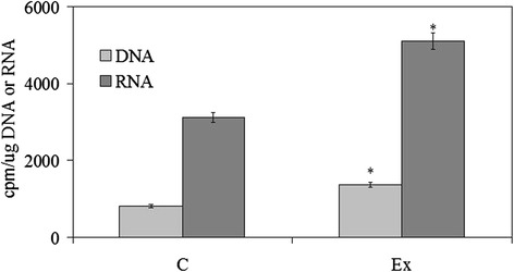
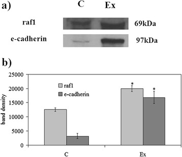
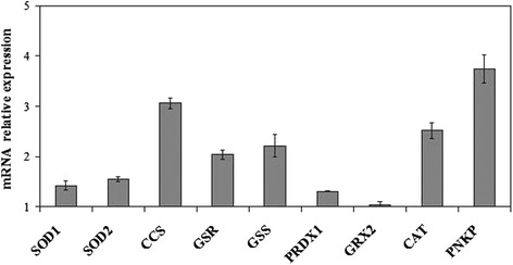
Results: We found that cholesterol changes cancer cell morphology with the appearance of protrusions together to the down expression of β-actin gene and reduction of β-actin protein. The lipid influences SUP-T1 cell aggressiveness since stimulates DNA and RNA synthesis for cell proliferation and increases raf1 and E-cadherin, molecules involved in invasion and migration of cancer cells. Cholesterol does not change GRX2 expression but it overexpresses SOD1, SOD2, CCS, PRDX1, GSR, GSS, CAT and PNKP. We suggest that cholesterol reaches the nucleus and increases the nuclear lipid microdomains known to act as platform for chromatin anchoring and gene expression.

Conclusion: The results imply that, in hypercholesterolemia conditions, cholesterol reaches the nuclear lipid microdomains where activates gene expression coding for antioxidant proteins. We propose the cholesterolemia as useful parameter to monitor in patients with cancer.


Figures

Fig. 1
Fig. 1
SUP-T1 morfology. The cells were cultured without (control) or with 800nM CHO (experimental) for three days, then they were treated as reported in Materials and Methods section
Fig. 2
Fig. 2
Effect of cholesterol on β-actin protein. Immunoblot of proteins was probed with anti-β-actin and visualized by ECL after 24 h of culture without (control) or with 800nM CHO (experimental). Apparent molecular weight (43 kDa) was calculated according to the migration of molecular size standards. The area density was calculated with Scion Image programme on densitometry scanning; the data represent the mean ± S.D. of three experiments performed in duplicate. (Significance, *P < 0.001 versus Control sample)
Fig. 3
Fig. 3
Effect of cholesterol on DNA and RNA synthesis. DNA synthesis was studied by evaluating the incorporation of 3H-thymidine in the DNA and RNA synthesis by evaluating the incorporation of 3H-UTP in the RNA. The specific activity was calculated as cpm/μg DNA and cpm/μg RNA, respectively. The data represent the mean ± S.D. of three experiments performed in duplicate. (Significance, *P < 0.001 versus Control sample)
Fig. 4
Fig. 4
Effect of cholesterol on raf1 and e-cadherin proteins. a Immunoblot of proteins was probed with specific antibodies and visualized by ECL after 24 h of culture without (control) or with 800nM CHO (experimental). Apparent molecular weights were calculated according to the migration of molecular size standards; b The area density was calculated with Scion Image programme on densitometry scanning; the data represent the mean ± S.D. of three experiments performed in duplicate. (Significance, *P < 0.001 versus Control sample)
Fig. 5
Fig. 5
Effect of cholesterol on SOD1, SOD2, CCS, GSR, GSS, PRDX1, GRX2, CAT, PNKP expression. RTqPCR analysis was performed in control (without CHO) and experimental SUP-T1 cells (with 800nM CHO) collected after 24 h of culture. The results were normalized with the levels of the GAPDH and expressed as mRNA of experimental sample versus control sample. Data are expressed as the mean ± S.D. of 3 independent experiments performed in three PCR replicates
Fig. 6
Fig. 6
SUP-T1 composition. The cells were cultured without (control) or with 800nM CHO (experimental) and collected after 24 h. a cholesterol (CHO), sphingomyelin (SM), DNA and RNA content in cells, nuclei (N) and nuclear lipid microdomains (NLM), the data are expressed as μg/106 cells and represent the mean ± S.D. of four experiments performed in duplicate; b CHO, SM, DNA and RNA content in NLM, the data are expressed as μg/mg protein and represent the mean ± S.D. of four experiments performed in duplicate. (Significance, *P < 0.001 versus Control sample)
Fig. 7
Fig. 7
Sphingomyelinase activity. The cells were cultured without (control) or with 800nM CHO (experimental) and collected after 24 h; cell, nuclei (N) and nuclear lipid microdomains (NLM) were prepared as reported in Materials and Methods section. a The data are expressed as pmol/mg protein/min; b the data are expressed as percentage respect to control samples. (Significance, *P < 0.001 versus Control sample)

References

    1. Moon H, Ruelcke JE, Choi E, Sharpe LJ, Nassar ZD, Bielefeldt-Ohmann H, et al. Diet-induced hypercholesterolemia promotes androgen-independent prostate cancer metastasis via IQGAP1 and caveolin-1. Oncotarget. 2015;6:7438–7453. doi: 10.18632/oncotarget.3476. - DOI - PMC - PubMed
    1. Hu J, La Vecchia C, Negri E, de Groh M, Morrison H, Mery L. Macronutrient intake and stomach cancer. Cancer Causes Control. 2015;26:839–847. doi: 10.1007/s10552-015-0557-9. - DOI - PubMed
    1. Pelton K, Coticchia CM, Curatolo AS, Schaffner CP, Zurakowski D, Solomon KR, et al. Hypercholesterolemia induces angiogenesis and accelerates growth of breast tumors in vivo. Am J Pathol. 2014;184:2099–2110. doi: 10.1016/j.ajpath.2014.03.006. - DOI - PMC - PubMed
    1. Llaverias G, Danilo C, Mercier I, Daumer K, Capozza F, Williams TM, et al. Role of Cholesterol in the Development and Progression of Breast Cancer. Am J Pathol. 2011;178:402–413. doi: 10.1016/j.ajpath.2010.11.005. - DOI - PMC - PubMed
    1. Iso H, Ikeda A, Inoue M, Sato S, Tsugane S. Serum cholesterol levels in relation to the incidence of cancer: the JPHC study cohorts. Int J Cancer. 2009;125:2679–2686. doi: 10.1002/ijc.24668. - DOI - PubMed

Publication types

MeSH terms