Review
. 2016 Apr;13(2):295-310.
doi: 10.1007/s13311-015-0415-1.

Electrical Stimulation to Enhance Axon Regeneration After Peripheral Nerve Injuries in Animal Models and Humans

Affiliations
Review

Electrical Stimulation to Enhance Axon Regeneration After Peripheral Nerve Injuries in Animal Models and Humans

Tessa Gordon. Neurotherapeutics. 2016 Apr.

Abstract

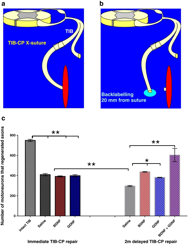
Injured peripheral nerves regenerate their lost axons but functional recovery in humans is frequently disappointing. This is so particularly when injuries require regeneration over long distances and/or over long time periods. Fat replacement of chronically denervated muscles, a commonly accepted explanation, does not account for poor functional recovery. Rather, the basis for the poor nerve regeneration is the transient expression of growth-associated genes that accounts for declining regenerative capacity of neurons and the regenerative support of Schwann cells over time. Brief low-frequency electrical stimulation accelerates motor and sensory axon outgrowth across injury sites that, even after delayed surgical repair of injured nerves in animal models and patients, enhances nerve regeneration and target reinnervation. The stimulation elevates neuronal cyclic adenosine monophosphate and, in turn, the expression of neurotrophic factors and other growth-associated genes, including cytoskeletal proteins. Electrical stimulation of denervated muscles immediately after nerve transection and surgical repair also accelerates muscle reinnervation but, at this time, how the daily requirement of long-duration electrical pulses can be delivered to muscles remains a practical issue prior to translation to patients. Finally, the technique of inserting autologous nerve grafts that bridge between a donor nerve and an adjacent recipient denervated nerve stump significantly improves nerve regeneration after delayed nerve repair, the donor nerves sustaining the capacity of the denervated Schwann cells to support nerve regeneration. These reviewed methods to promote nerve regeneration and, in turn, to enhance functional recovery after nerve injury and surgical repair are sufficiently promising for early translation to the clinic.

Keywords: Delayed nerve repair; Electrical stimulation; Peripheral nerve injury; Peripheral nerve regeneration; Side-to-side crossbridges.


Conflict of interest statement

Compliance with Ethical Standards Required Author Forms Disclosure forms provided by the authors are available with the online version of this article.

Figures

Fig. 1
Fig. 1
Outgrowth of regenerating axons from a common peroneal nerve sutured directly to denervated soleus muscle. Silver nitrate-stained axons grow out from the suture site (at the bottom of the micrograph) and follow the muscle fibers to reinnervate many denervated endplates that are identified by their immunostaining with cholinesterase (blue)
Fig. 2
Fig. 2
Progressive failure of regenerating axons to grow through an autograft into the distal nerve stump and to reinnervate denervated muscles after delayed nerve repair. (a) The experimental protocol was to tease Sprague–Dawley rat ventral roots to evoke and record isometric contractile force from muscle fibers in the reinnervated tibialis anterior (TA) muscle that were reinnervated by single tibial (Tib) axons (motor units) at least 6 months after cross-suturing the proximal tibial (Tibp) nerve stump to the distal common peroneal (CPd) nerve via a CP nerve autograft (CPg) taken from the contralateral hindlimb. The cross-suture took place either (a) immediately or after a delay of up to 500 days, resulting in (b) chronic TIB axotomy, (c) chronic CP graft denervation, or (d) chronic TA muscle denervation. The relative contributions of (b) chronic axotomy, (c) chronic CP nerve denervation, and (d) TA muscle denervation to the decline in the number of regenerating Tib nerves that reinnervated the denervated TA muscle (motor unit number) are seen with each contributing to the progressive failure of nerve regeneration
Fig. 3
Fig. 3
The transient expression of regeneration-associated genes in injured neurons after transecting the peripheral nerve. (a) The chromatolytic reaction of an injured motoneuron includes the movement of the nucleus to an eccentric position with expression of several growth-associated genes (GAGs) that include the neurotrophic factors and their receptors. There is a switch from the transmitting to the growth state in the axotomized motoneurons that includes the downregulation of transmitter associated genes (choline acetyltransferase and acetylcholinesterase) and the cytoskeletal protein, neurofilament, concurrent with a decline in the size of the axons proximal to the lesion [38]. The cytoskeletal proteins of tubulin and actin are upregulated, as are several growth-associated proteins (GAPs) that include GAP-43. However, this expression is transient as shown by the examples of the decline in the expression of (b) tubulin in chronically axotomized sciatic motoneurons, (c) glial-derived neurotrophic factor (GDNF), and the p75 receptor for the neurotrophic factors in chronically denervated Schwann cells. Mean ± SE bars are plotted in the graphs and the data points are fitted by exponential lines
Fig. 4
Fig. 4
Brief 20-Hz electrical stimulation accelerates axon outgrowth across the surgical site of nerve repair. (a) The rat femoral nerve was transected and microsurgically repaired 20 mm from the branching of the nerves into the motor branch to the quadriceps muscle and the cutaneous saphenous nerve branch. The number of backlabeled motoneurons that regenerated their axons into both the branches and were backlabeled 25 mm from the suture site with fluororuby and fluorogold, increased progressively over an 8–10-week period of time. (b) Staggered axon regeneration was due to the “staggering” of regenerating axons across the suture site over a 4-week period as determined by counting motoneurons that were backlabeled by crushing the femoral nerve stump 1.5 mm from the repair site. (c, d) The “staggered axon regeneration” across the suture site was accelerated significantly (*p < 0.05) by a 1-h period of 20-Hz continuous electrical stimulation such that all motoneurons regenerated their axons over a distance of 25 mm within 3 weeks rather than 8-10 weeks with sham stimulation and motoneurons regenerated their axons significantly more qucikly across the suture site into the distal nerve stump
Fig. 5
Fig. 5
Brief electrical stimulation accelerates axon outgrowth across the nerve injury site after both immediate and delayed nerve repair. Two months after cross-suture of the common peroneal (CP) proximal nerve stump and the tibial (TIB) distal nerve stump, the numbers of backlabeled CP motor and sensory neurons (left and right set of histograms, respectively) that regenerated their axons 10 mm into the TIB nerve stump was increased significantly (*p < 0.05) when the CP nerve proximal to the repair site was subjected to a 1-h period of electrical stimulation after (a) immediate nerve repair, (b) 2 months (2 m) chronic axotomy of the CP neurons, (c) 2 months after chronic denervation of the TIB distal nerve stump, and (d) after 3 months chronic axotomy and denervation of the CP neurons and the distal TIB nerve stump. SC = Schwann cells
Fig. 6
Fig. 6
Brief electrical stimulation accelerates reinnervation of muscles after carpal tunnel (CT) release surgery in humans with severe CT syndrome. (a) Electromyographic (EMG) signals were recorded from surface electrodes on the thenar eminence in response to maximal and all-or-none stimulation of the median nerve. (b) The ratio of the compound action potential (CMAP) and mean motor unit potential (MUAP) provided the motor unit number estimation. Examples of MUAPs recorded in response to progressive movement of the stimulating electrodes up the arm [in (a)] are shown in (b) with the CMAP. (c) The standard errors of the mean values before CT release (shown as dotted lines) of motor unit number before the CT release surgery demonstrate that ~50 % of median motoneurons were axotomized by pressure exerted by the ligament that overlies the nerve at the wrist. When the median nerve was not stimulated after the CT release surgery, the trend to increase above the preoperative number of motor units was not significant. In contrast, in those patients whose median nerve was electrically stimulated for 1 h continuously at 20 Hz, the entire thenar muscle was reinnervated with restoration of the normal number of motor units (shown as an interrupted line)
Fig. 7
Fig. 7
Rolipram, a phosphodiesterase that increases intracellular cyclic adenosine monophosphate (cAMP) in neurons or brief electrical stimulation accelerates axon outgrowth across the suture line of a transected and surgically repaired peripheral nerve in rats. (a) Rolipram administered locally to a surgically repaired common peroneal nerve or (b) 1 h continuous 20-Hz electrical stimulation proximal to the surgical repair site of a transected femoral nerve significantly increases the number of motoneurons that regenerate their axons. (c) Protein kinase A is activated by cAMP to promote transcription of proteins that include the neurotrophic factors that amplify the effects of cAMP in promoting gene transcription of growth associated genes such as tubulin and actin and, in turn, accelerate axon outgrowth (p < 0.05). BDNF = brain-derived neurotrophic factor; ATP = adenosine triphosphate; PKA = protein kinase A
Fig. 8
Fig. 8
A conditioning lesion and brief electrical stimulation (ES) promotes nerve regeneration within the spinal cord. (ia) Following a spinal lesion at the level of T8, few axons entered the lesion site [camara lucida drawings with example of cholerotoxin (CTX)-stained axons 1 week after injecting CTX into the sciatic nerve] after sham electrical stimulation at 20 Hz for 1 h (or a sham conditional lesion, not shown). (ib) In contrast, a conditioning sciatic crush lesion (CL) resulted in more regeneration of CTX-labeled axons, with (ic) an intermediate number regenerating after 1 h of 20-Hz continuous electrical stimulation of the sciatic nerve. (iia) The significantly (p < 0.05) elevated number of regenerated sciatic nerve axons that grew out and over longer distances from the lesion site after a CL and (iib) the significantly elevated numbers of axons that grew out from the lesion site but did not grow over longer distances after 20Hz electrical stimulation for 1 h. Each point is the mean ± SEM
Fig. 9
Fig. 9
Exogenous administration of the neurotrophic factors brain-derived neurotrophic factor (BDNF) and glial cell-line derived neurotrophic factor (GDNF) promotes neuronal regeneration of injured motoneurons after delayed nerve repair. (a) A cross-suture (X-suture) technique to cross-suture the freshly cut tibial (TIB) proximal nerve stump to the distal stump of the freshly cut common peroneal (CP) nerve or to cross-suture the TIB proximal nerve stump that was ligated for 2 months (2 m) previously (to prolong TIB motoneuron axotomy) to a freshly cut CP distal nerve stump. The neurotrophic factors were delivered from a miniosmotic pump to the coaptation site over a 1-month period. (b) Thereafter, the regenerated TIB axons in the CP nerve were exposed to fluorogold to backlabel those TIB motoneurons that regenerated their axons over a 20-mm distance. (c) Saline-treated axons regenerated ~45 % of their axons within the a month of regeneration, consistent with the staggered regeneration demonstrated by Al-Majed et al. [6]. This regeneration was significantly reduced by a 2-month period of chronic axotomy consistent with the data of Fu and Gordon [17]. The decline in motoneuron numbers after 2 months was not only prevented by a month of administration of BDNF (2 μg/day) or GDNF (0.1 μg/day), but also the neurotrophic factors significantly increased the numbers of 2-month chronically axotomized motoneurons that regenerated their axons (* p < 0.05; ** p < 0.01)

References

    1. Fenrich K, Gordon T. Canadian Association of Neuroscience review: axonal regeneration in the peripheral and central nervous systems-current issues and advances. Neurol Sci 2004;31:142--156. - PubMed
    1. Fu SY, Gordon T. The cellular and molecular basis of peripheral nerve regeneration. Mol Neurobiol. 1997;14:67–116. doi: 10.1007/BF02740621. - DOI - PubMed
    1. Brushart TM, Hoffman PN, Royall RM, Murinson BB, Witzel C, Gordon T. Electrical stimulation promotes motoneuron regeneration without increasing its speed or conditioning the neuron. J Neurosci. 2002;22:6631–38. - PMC - PubMed
    1. Al-Majed AA, Neumann CM, Brushart TM, Gordon T. Brief electrical stimulation promotes the speed and accuracy of motor axonal regeneration. J Neurosci. 2000;20:2602–2608. - PMC - PubMed
    1. Elzinga K, Tyreman N, Ladak A, Savaryn B, Olson J, Gordon T. Brief electrical stimulation improves nerve regeneration after delayed repair in Sprague Dawley rats. Exp Neurol. 2015;269:142–153. doi: 10.1016/j.expneurol.2015.03.022. - DOI - PubMed

Publication types