Review
. 2016 Feb;18(1):1-17.
doi: 10.1007/s11307-015-0919-4.

Site-Specifically Labeled Immunoconjugates for Molecular Imaging--Part 1: Cysteine Residues and Glycans

Affiliations
Review

Site-Specifically Labeled Immunoconjugates for Molecular Imaging--Part 1: Cysteine Residues and Glycans

Pierre Adumeau et al. Mol Imaging Biol. 2016 Feb.

Abstract

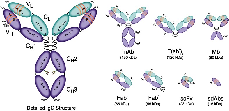
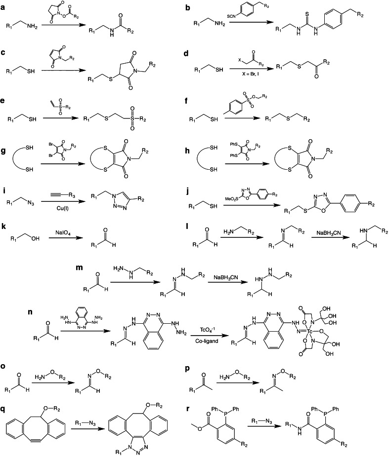
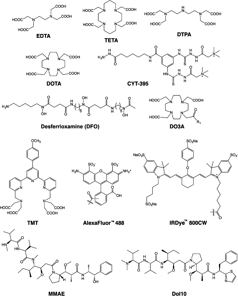
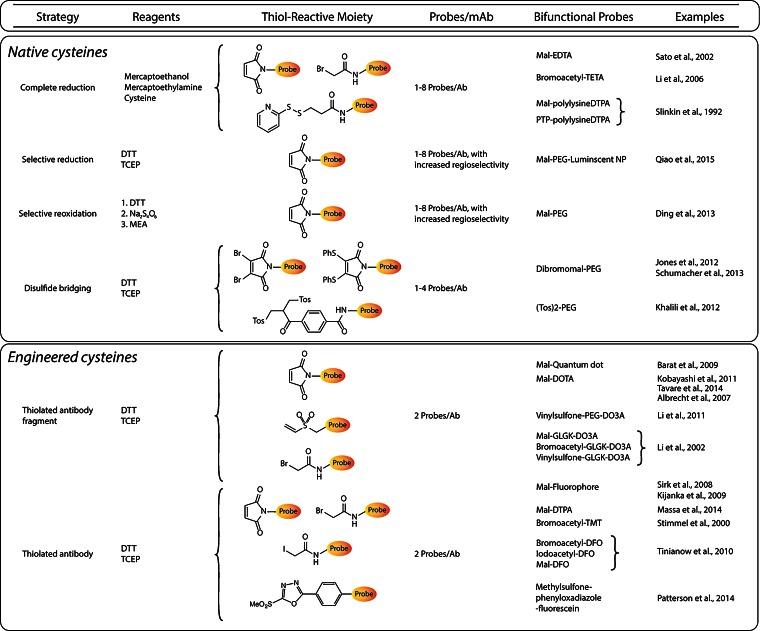
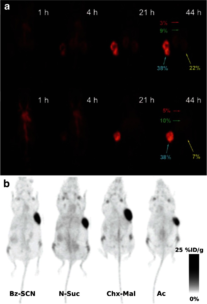
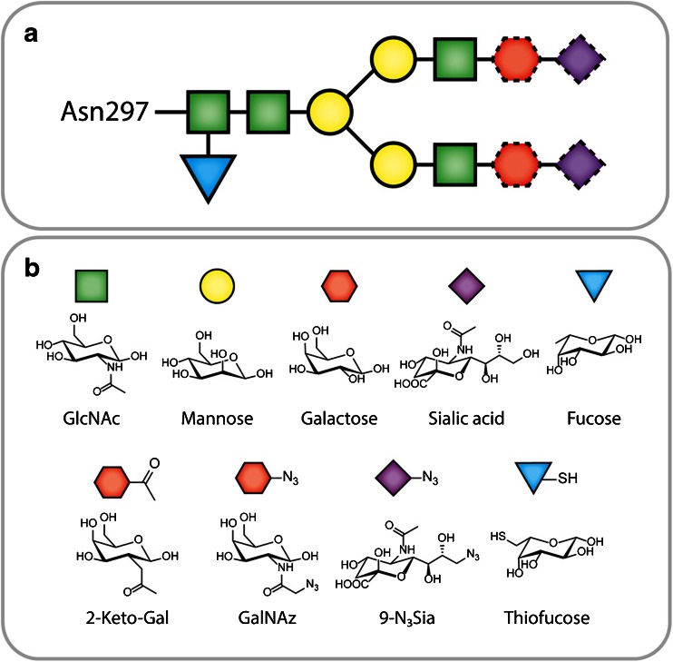
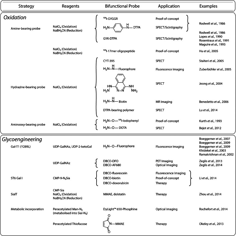
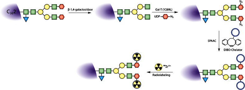
Due to their remarkable selectivity and specificity for cancer biomarkers, immunoconjugates have emerged as extremely promising vectors for the delivery of diagnostic radioisotopes and fluorophores to malignant tissues. Paradoxically, however, these tools for precision medicine are synthesized in a remarkably imprecise way. Indeed, the vast majority of immunoconjugates are created via the random conjugation of bifunctional probes (e.g., DOTA-NCS) to amino acids within the antibody (e.g., lysines). Yet antibodies have multiple copies of these residues throughout their macromolecular structure, making control over the location of the conjugation reaction impossible. This lack of site specificity can lead to the formation of poorly defined, heterogeneous immunoconjugates with suboptimal in vivo behavior. Over the past decade, interest in the synthesis and development of site-specifically labeled immunoconjugates--both antibody-drug conjugates as well as constructs for in vivo imaging--has increased dramatically, and a number of reports have suggested that these better defined, more homogeneous constructs exhibit improved performance in vivo compared to their randomly modified cousins. In this two-part review, we seek to provide an overview of the various methods that have been developed to create site-specifically modified immunoconjugates for positron emission tomography, single photon emission computed tomography, and fluorescence imaging. We will begin with an introduction to the structure of antibodies and antibody fragments. This is followed by the core of the work: sections detailing the four different approaches to site-specific modification strategies based on cysteine residues, glycans, peptide tags, and unnatural amino acids. These discussions will be divided into two installments: cysteine residues and glycans will be detailed in Part 1 of the review, while peptide tags and unnatural amino acids will be addressed in Part 2. Ultimately, we sincerely hope that this review fosters interest and enthusiasm for site-specific immunoconjugates within the nuclear medicine and molecular imaging communities.

Keywords: Antibody; Antibody fragment; Bioconjugation; Bioorthogonal chemistry; Click chemistry; Cysteine; Fluorescence imaging; Glycans; Glycoengineering; Immunoglobulins; Maleimide; Near-infrared fluorescence imaging; Optical imaging; PET; Positron emission tomography; Protein engineering; SPECT; Single photon emission computed tomography; Site-selective conjugation; Site-specific conjugation.


Figures

Fig. 1
Fig. 1
Detailed structural schematic of a full-length IgG as well as an assortment of antibody fragments.
Fig. 2
Fig. 2
The basic chemical reactions underpinning the bioconjugation strategies discussed in this work.
Fig. 3
Fig. 3
Selected chelators and cargoes used in the site-specifically labeled immunoconjugates discussed in this work.
Fig. 4
Fig. 4
Table of site-specific bioconjugation strategies based on the modification of cysteine residues.
Fig. 5
Fig. 5
a Serial PET images of site-specifically labeled [64Cu]DOTA-PEG24-AVP04-50 (top) and [64Cu]DOTA-PEG48-AVP04-50 (bottom) in athymic nude mice bearing LS174T xenografts. The labels in red, green, yellow, and turquoise illustrate the %ID/g values in the heart, liver, kidney, and tumor, respectively. Figure adapted and reprinted with the permission of Li et al. Copyright 2011 American Chemical Society [65]. b PET images of four different variants of [89Zr]DFO-thio-trastuzumab in mice bearing BT474 xenografts. In two of the radioimmunoconjugates, the chelator was attached using nonsite-specific conjugation methods (Bz-SCN and N-Suc), while in the other two constructs, bioconjugation was achieved using thiol-reactive variants of DFO (Chx-Mal and Ac). Figure adapted and reprinted with the permission of Tinianow et al. Copyright 2010 Elsevier Publishing Group [17].
Fig. 6
Fig. 6
a The biantennary structure of the heavy chain glycans; the dotted outlines indicate residues that are not always present in the glycans. b Structures of natural and synthetic monosaccharides.
Fig. 7
Fig. 7
Site-specific bioconjugation strategies based on the modification of glycans.
Fig. 8
Fig. 8
Schematic of a Gal-T(Y289L)-based site-specific modification procedure.
Fig. 9
Fig. 9
a PET and b near-infrared fluorescence images of athymic nude mice bearing SW1222 tumors (white arrows) injected with either site-specifically labeled or traditionally labeled [89Zr]DFO-huA33-Alexa Fluor® 680. In the PET images, the coronal slices intersect the center of the tumors. Figure adapted and reprinted with the permission of Zeglis et al. Copyright 2014 American Chemical Society [18].

References

    1. Wu AM. Antibodies and antimatter: the resurgence of immuno-PET. J Nucl Med. 2009;50:2–5. doi: 10.2967/jnumed.108.056887. - DOI - PubMed
    1. van Dongen GAMS, Visser GWM, Lub-de Hooge MN, de Vries EG, Perk LR. Immuno-PET: a navigator in monoclonal antibody development and applications. Oncologist. 2007;12:1379–1389. doi: 10.1634/theoncologist.12-12-1379. - DOI - PubMed
    1. Deri MA, Zeglis BM, Francesconi LC, Lewis JS. PET imaging with 89Zr: from radiochemistry to the clinic. Nucl Med Biol. 2013;40:3–14. doi: 10.1016/j.nucmedbio.2012.08.004. - DOI - PMC - PubMed
    1. Frangioni JV. In vivo near-infrared fluorescence imaging. Curr Opin Chem Biol. 2003;7:626–634. doi: 10.1016/j.cbpa.2003.08.007. - DOI - PubMed
    1. Cohen R, Vugts DJ, Stigter-van Walsum M, Visser GWM, van Dongen G. Inert coupling of IRDye800CW and zirconium-89 to monoclonal antibodies for single- or dual-mode fluorescence and PET imaging. Nat Protoc. 2013;8:1010–1018. doi: 10.1038/nprot.2013.054. - DOI - PubMed

Publication types