. 2016 Jul;73(13):2543-63.
doi: 10.1007/s00018-015-2128-3. Epub 2016 Jan 11.

Enhanced genome editing in mammalian cells with a modified dual-fluorescent surrogate system

Affiliations

Enhanced genome editing in mammalian cells with a modified dual-fluorescent surrogate system

Yan Zhou et al. Cell Mol Life Sci. 2016 Jul.

Abstract

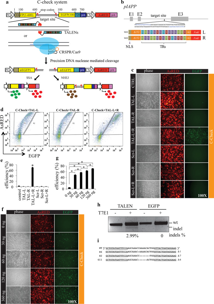
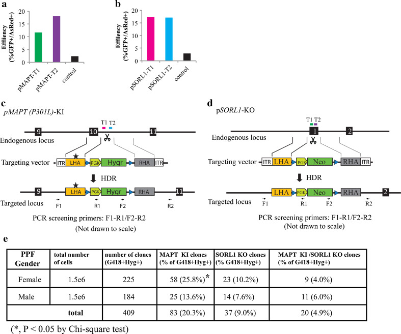
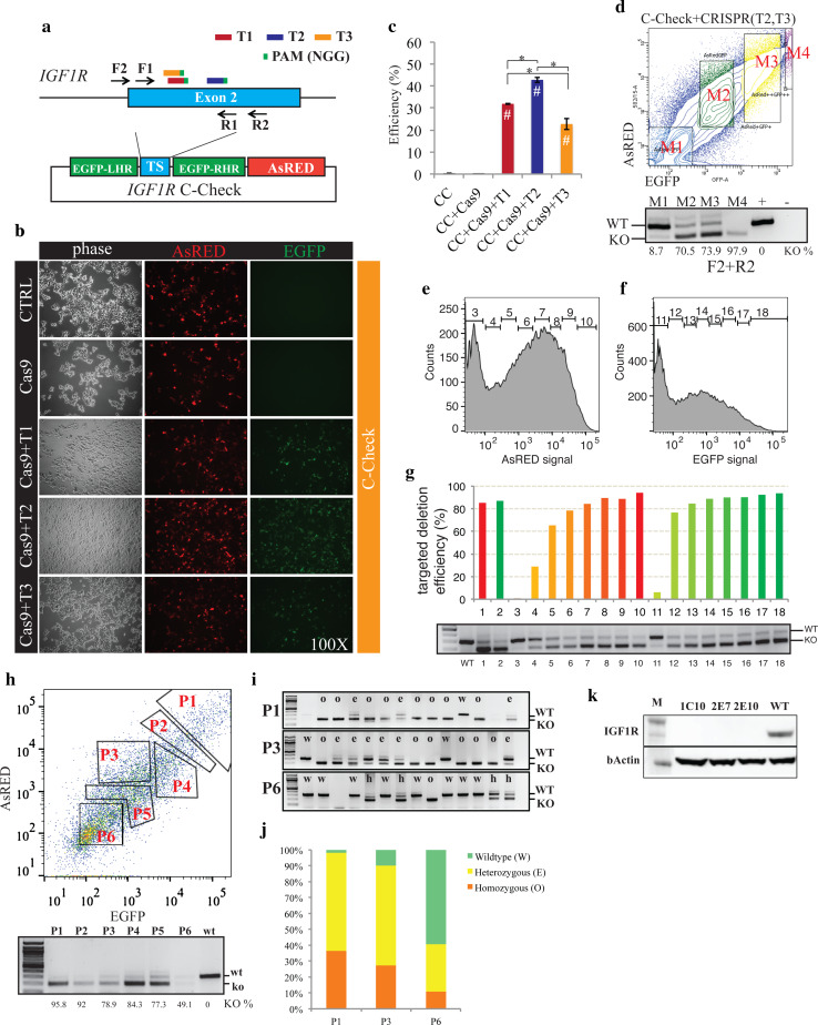
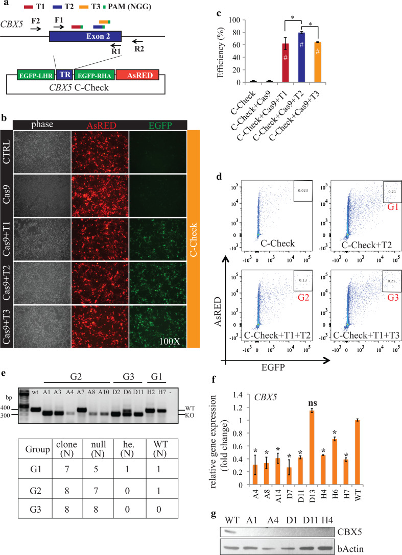
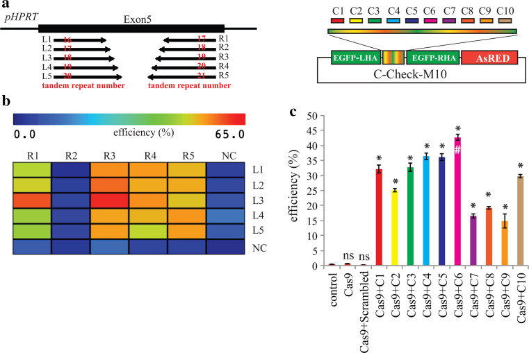
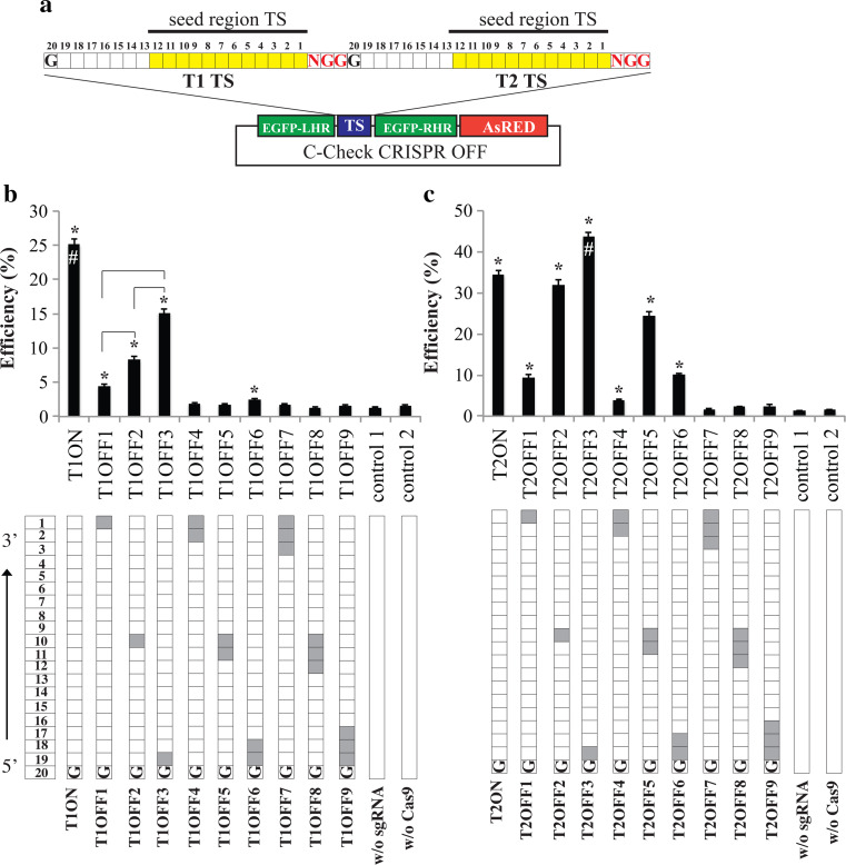
Programmable DNA nucleases such as TALENs and CRISPR/Cas9 are emerging as powerful tools for genome editing. Dual-fluorescent surrogate systems have been demonstrated by several studies to recapitulate DNA nuclease activity and enrich for genetically edited cells. In this study, we created a single-strand annealing-directed, dual-fluorescent surrogate reporter system, referred to as C-Check. We opted for the Golden Gate Cloning strategy to simplify C-Check construction. To demonstrate the utility of the C-Check system, we used the C-Check in combination with TALENs or CRISPR/Cas9 in different scenarios of gene editing experiments. First, we disrupted the endogenous pIAPP gene (3.0 % efficiency) by C-Check-validated TALENs in primary porcine fibroblasts (PPFs). Next, we achieved gene-editing efficiencies of 9.0-20.3 and 4.9 % when performing single- and double-gene targeting (MAPT and SORL1), respectively, in PPFs using C-Check-validated CRISPR/Cas9 vectors. Third, fluorescent tagging of endogenous genes (MYH6 and COL2A1, up to 10.0 % frequency) was achieved in human fibroblasts with C-Check-validated CRISPR/Cas9 vectors. We further demonstrated that the C-Check system could be applied to enrich for IGF1R null HEK293T cells and CBX5 null MCF-7 cells with frequencies of nearly 100.0 and 86.9 %, respectively. Most importantly, we further showed that the C-Check system is compatible with multiplexing and for studying CRISPR/Cas9 sgRNA specificity. The C-Check system may serve as an alternative dual-fluorescent surrogate tool for measuring DNA nuclease activity and enrichment of gene-edited cells, and may thereby aid in streamlining programmable DNA nuclease-mediated genome editing and biological research.

Keywords: CRISPR/Cas9; Dual-fluorescent surrogate reporter; Gene targeting; Genome engineering; Homologous recombination; Single-strand annealing; TALENs.


Conflict of interest statement

YL (2012–2014), SGR, and HD were financed by Novo Nordisk A/S. TK was financed by Gubra ApS. A patent claim is declared to the generation of a diabetes pig model based on genetic modification of the porcine IAPP gene. No other competing interests are declared by the authors.

Figures

Fig. 1
Fig. 1
Generation of the C-Check system and validation for functional assay of TALEN-mediated DNA cleavage activity. a Schematic illustration of the C-Check reporter system. PGK phosphoglycerate kinase 1 promoter; the coding sequence of the EGFP gene is indicated with codon numbering from 5′ to 3′; the homology arms within the two truncated EGFP fragments (trEGFP1 and trEGFP2) are indicated by the yellow boxes; different poly A terminal signals were used to avoid recombination as indicated in different color. Binding of TALENs and CRISPR/Cas9 to the target sites in the C-Check reporter vector is illustrated. After cleavage of the episomal C-Check reporter vector in cells, the C-Check vector can be repaired through two pathways: single-strand annealing (SSA) or non-homologous end joining (NHEJ). Two stop codons were included to flank the target sites in the C-Check vector. The first stop codon was pre-built in the 5′-end and the second stop codon at the 3′-end is introduced by Golden Gate insertion of the target site sequence. b Schematic representation of the porcine IAPP locus and IAPP target site. Exons are indicated with gray boxes and the target site sequences are highlighted in blue. NLS nuclear localization signal, TRs tandem repeats, L and R denote the TALEN monomer protein that binds to the target site at the coding and non-coding strand. Figures are not drawn to scale. c Representative fluorescence imaging of the C-Check assaying of IAPP TALENs activity. Scr scrambled TALENs that do not target IAPP. d Representative flow cytometry diagram of the nuclease activity quantification by C-Check. Weak transmission from the AsRED spectrum to the EGFP detector was observed. The indicated gating (P1 and P2) was applied to avoid any false positive results. Efficiency was calculated as the percentage of cells in P2 out of the total number of cells in P1 and P2. This gating and quantification strategy was applied to all C-Check nuclease quantification assays throughout the study. e Quantification of IAPP TALEN activity. Asterisk (*) indicates a p value less than 0.05 compared to the remaining groups. f, g Representative fluorescence images and quantification of dose-dependent TALEN nuclease activities determined by C-Check analysis. Asterisk (*) indicates a p value less than 0.05 between the compared groups. h T7E1 assay of IAPP TALEN-induced indels in primary porcine fibroblasts. i Identification of TALEN-induced indels by Sanger sequencing. Three out of 96 clones analyzed carried 1, 7, and 6 bp deletions at the TALEN spacer sites, respectively. TALEN target sites are underlined
Fig. 2
Fig. 2
Double-gene targeting in primary porcine fibroblasts with C-Check-validated CRISPR/Cas9. Quantification of MAPT (a) and SORL1 (b) CRISPR/Cas9 sgRNA activity by C-Check. HEK293T cells were co-transfected with the C-Check vector alone (control) or in different combinations of each sgRNA and the Cas9 vector. Cells were harvested 48 h post transfection and subjected to flow cytometry analysis. Schematic illustration of CRISPR/Cas9-mediated porcine MAPT (P301L) knockin (KI) by HDR (c) and CRISPR/Cas9-mediated pSORL1 knockout (KO) by HDR (d). Exons for each gene are indicated by black boxes. The CRISPR sgRNA target sites (pMAPT-T1, pMAPT-T2, pSORL1-T1, and pSORL1-T2) are indicated by a red, light blue, green, dark blue box, respectively. An asterisk (*) indicates the MAPT (P301L) mutation in the targeting vector and the targeted locus. Blue triangles denote LoxP sites. ITR inverted terminal repeats in the rAAV targeting plasmid, LHA and RHA left and right homology arm, respectively, Hygr hygromycin antibiotic resistance gene driven by a PGK promoter, Neo neomycin antibiotic resistance gene driven by a PGK promoter. Primers for PCR screening are indicated by arrows. Figures are not drawn to scale. e Summary of single- and double-gene targeting frequency using pMAPT-T2 and pSORL1-T1 in primary porcine fibroblasts (PPF)
Fig. 3
Fig. 3
Fluorescence tagging of MYH6 and COL2A1 in human fibroblasts. a Schematic representation of the CRISPR/Cas9-mediated C-terminal fluorescence tagging of endogenous genes by homologous recombination. One and two sgRNAs were generated for MYH6 and COL2A1, respectively, indicated with a correspondingly colored box. UTR, TS, LHA, RHA, and 2A denote untranslated region, target site, left homology arm, right homology arm, and 2A peptide, respectively. TK thymidine kinase cassette for Cre-mediated excision screening, PGK-Neo PGK promoter-driven neomycin expression cassette for gene targeting selection; two LoxP sites were included for excision of the antibiotic markers by Cre recombinase. b Schematic illustration of the Golden Gate assembly of the C-terminal EGFP-tagging system. “Kan” and “amp” denote bacterial kanamycin and ampicillin selection cassettes. The 2A peptide sequences are generated upon the correct assembly of the PGK-Neo-1A fragment and the 1A-TK fragment. c, d Quantitative analysis of MYH6 and COL2A1 sgRNA activity by C-Check assays. Asterisk (*) indicates statistical significance compared to the control (C-Check only); hash symbol indicates statistical significance compared to the Cas9 + COL2A1-T1. e, f Screening PCR of MYH6 and COL2A1 knockin of 10 and 13 G418+ clones. Symbols (P, +, −) indicate targeted knockin clones, positive control, and negative control templates, respectively
Fig. 4
Fig. 4
Enrichment of IGF1R null-modified HEK293T cells with the C-Check surrogate reporter vector. a Schematic illustration of the endogenous IGF1R locus and the IGF1R C-Check vector. All sgRNAs target sites (T1–T3) were on the coding strand of exon 2. Primers for generating the IGF1R C-Check vector (F1 + R1) and for screening of IGF1R knockout (F2 + R2) are indicated with black arrows. b, c Representative fluorescence imaging and quantification of sgRNAs activity by C-Check. Asterisk (*) indicates statistical significance between the comparisons; hash symbol indicates statistical significance compared to CC (transfected with the IGF1R C-Check plasmid only). d C-Check surrogate reporter-based FACS (upper panel) and PCR quantification of targeted IGF1R deletion frequency (KO%) in the indicated population of cells. The HEK93T cells were transfected with the IGF1R CRISPR/Cas9 (T2 and T3) and IGF1R C-Check (lower panel). Representative plot of AsRED-based (e) or EGFP-based (f) FACS sorting of HEK293T cells co-transfected with the IGF1R CRISPR/Cas9 (T2 and T3) and either a scrambled C-Check vecor (e) or the IGF1R C-Check vector (f). Gatings are illustrated with numbers (3–18). g Quantification of targeted IGF1R deletion efficiency based on PCR screening and Image J. Groups (3–18) are the corresponding sorted cells. Group 1 and 2 are unsorted cells co-transfected with the IGF1R CRISPR/Cas9 (T2 and T3) and either a scrambled C-Check vector or the IGF1R C-Check vector, respectively. Wild-type (WT) cells were used as control. h The HEK293T cells co-transfected with the IGF1R CRISPR/Cas9 (T2 and T3) and the IGF1R C-Check vector and sorted into six populations based on both AsRED and EGFP signal. The IGF1R knockout efficiency was quantified by PCR and image J (h, lower panel). i, j The cell populations (h, P1, P3, and P6) were also single-cell sorted into a 96-well plate for clonogenic cell growth followed by IGF1R knockout PCR screening of clonogenic cell clones. Letters o, e, and w represent homozygous, heterozygous, and “wild type” clones, respectively, based on PCR. Note that small indels could not be distinguished by PCR. The “wild type” bands appearing in the heterozygous and wild-type clones might therefore actually be mutated. This was further validated by Sanger sequencing (Additional File 6). k Western blot analysis of IGF1R in three IGF1R knockout cell clones. Wild-type (WT) parental HEK293T cells were used as control. Beta-actin was used as loading control
Fig. 5
Fig. 5
Enrichment of CBX5 null MCF-7 cells with the C-Check surrogate reporter vector. a Schematic illustration of the endogenous CBX5 locus and the CBX5 C-Check vector. All sgRNA target sites (T1–T3) were on the coding strand of CBX5 exon 2. Primers for generating the CBX5 C-Check vector (F1 + R1) and for screening of CBX5 knockout (F2 + R2) are denoted with black arrows. b, c Representative fluorescence imaging and quantification of sgRNAs activity by C-Check. Asterisk (*) indicates statistical significance between the corresponding comparisons; hash symbol indicates statistical significance compared to C-Check control (transfected with CBX5 C-Check plasmid only). d Illustration of FACS diagram and C-Check based gating for single-cells sorting. G1, G2, and G3 samples were population sorted into 96-well plates as described in the method section. e Genotyping by PCR and Sanger sequencing of CBX5 knockout clonogenic MCF-7 cells resulting from single-cell sorting. A summary of all clones genotyped by PCR and Sanger Sequencing is provided in the lower panel (Additional File 7). Note that small indels could not be distinguished by PCR screening. null targeted modified in all alleles, he. heterozygously modified. f qPCR analysis of 9 biallelic CBX5 knockout clones. Asterisk (*) indicates statistical significance compared to wild-type cells (WT); ns not significant. g Western blot analysis of CBX5 in five CBX5 knockout cell clones. Wild-type (WT) parental MCF-7 cells were used as control. Beta-actin was used as loading control
Fig. 6
Fig. 6
C-Check system is compatible with multiplexing nuclease analyses. a Schematic representation of the porcine HPRT locus (exon 5 and partial flanking introns), and the HPRT TALEN target sites (left panel) and the multiplex C-Check vector containing ten CRISPR/Cas9 targeting sites (right panel). Numbers of tandem repeats for each TALEN monomer protein is given with numbers in red. b Heatmap presentation of the TALEN nuclease activities for 25 pairs of HPRT TALENs. c Quantification of activity of ten CRISPR/Cas9 vectors by flow cytometry using a single multiplexing C-Check vector. Asterisk (*) indicates statistical significance compared to control (C-Check only); ns not significant compared to control; hash symbol indicates statistical significance compared to all other sgRNAs
Fig. 7
Fig. 7
Quantification of CRISPR/Cas9 sgRNA specificity with C-Check. a Illustration of the C-Check CRISPR OFF vector. Two sgRNA target sites were cloned into the C-Check vector. Positions for each nucleotide, represented with an individual box, in the protospacer sequence are annotated as 1–20 from the 3′-end to the 5′-end. Position 1 is the nucleotide preceding the PAM. The seed region of the target site (TS, 1–12) is colored yellow. b, c Quantification of CRISPR/Cas9 nuclease activity of one on-target (ON) sgRNA and nine off-target (OFF) sgRNAs for target site T1 (b) and T2 (c). The filled boxes in the lower panels represent mismatches between the sgRNA and the target site. Asterisk (*) indicates statistical significance compared to both controls; hash symbol, represents statistical significance compared to all remaining groups

References

    1. Gaj T, Guo J, Kato Y, Sirk SJ, Barbas CF., 3rd Targeted gene knockout by direct delivery of zinc-finger nuclease proteins. Nat Methods. 2012;9:805–807. doi: 10.1038/nmeth.2030. - DOI - PMC - PubMed
    1. Kim YG, Chandrasegaran S. Chimeric restriction endonuclease. Proc Natl Acad Sci USA. 1994;91:883–887. doi: 10.1073/pnas.91.3.883. - DOI - PMC - PubMed
    1. Hockemeyer D, Soldner F, Beard C, Gao Q, Mitalipova M, DeKelver RC, Katibah GE, Amora R, Boydston EA, Zeitler B, et al. Efficient targeting of expressed and silent genes in human ESCs and iPSCs using zinc-finger nucleases. Nat Biotechnol. 2009;27:851–857. doi: 10.1038/nbt.1562. - DOI - PMC - PubMed
    1. Hockemeyer D, Wang H, Kiani S, Lai CS, Gao Q, Cassady JP, Cost GJ, Zhang L, Santiago Y, Miller JC, et al. Genetic engineering of human pluripotent cells using TALE nucleases. Nat Biotechnol. 2011;29:731–734. doi: 10.1038/nbt.1927. - DOI - PMC - PubMed
    1. Bogdanove AJ, Voytas DF. TAL effectors: customizable proteins for DNA targeting. Science. 2011;333:1843–1846. doi: 10.1126/science.1204094. - DOI - PubMed

Publication types

MeSH terms

Substances