. 2016 Jan 11;5(1):e002555.
doi: 10.1161/JAHA.115.002555.

Mitochondrial Reactive Oxygen Species Mediate Cardiac Structural, Functional, and Mitochondrial Consequences of Diet-Induced Metabolic Heart Disease

Affiliations

Mitochondrial Reactive Oxygen Species Mediate Cardiac Structural, Functional, and Mitochondrial Consequences of Diet-Induced Metabolic Heart Disease

Aaron L Sverdlov et al. J Am Heart Assoc. .

Abstract

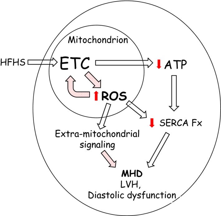
Background: Mitochondrial reactive oxygen species (ROS) are associated with metabolic heart disease (MHD). However, the mechanism by which ROS cause MHD is unknown. We tested the hypothesis that mitochondrial ROS are a key mediator of MHD.

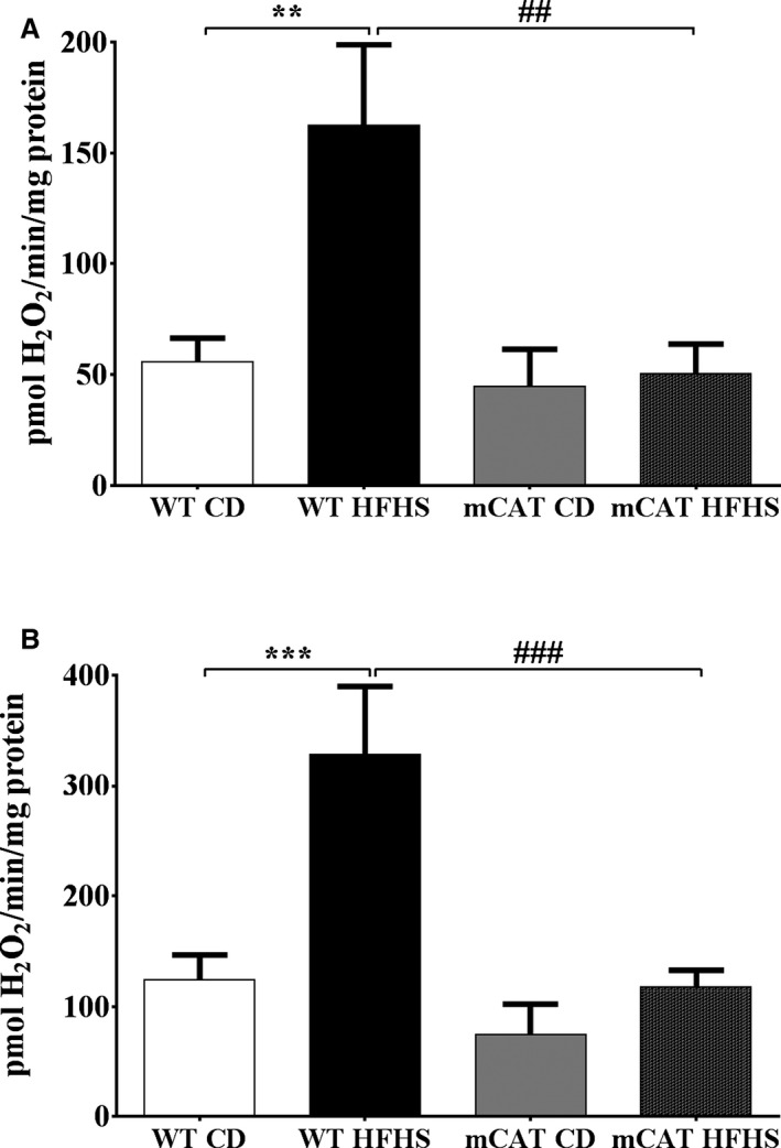
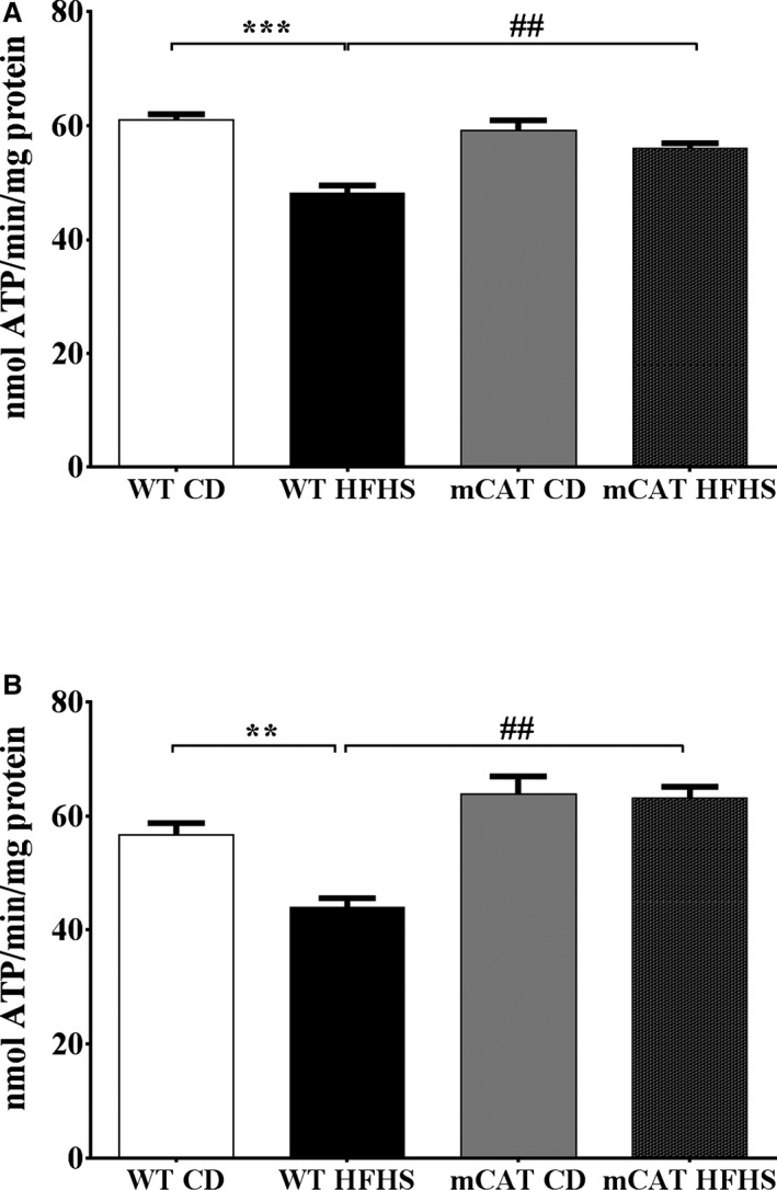
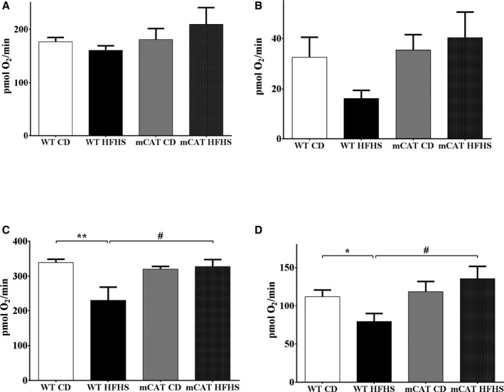
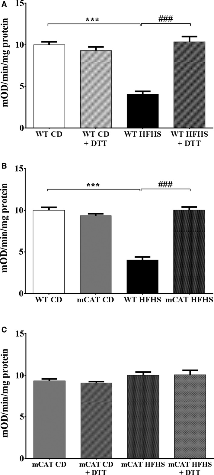
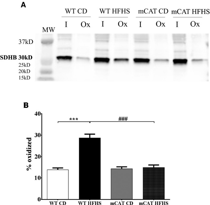
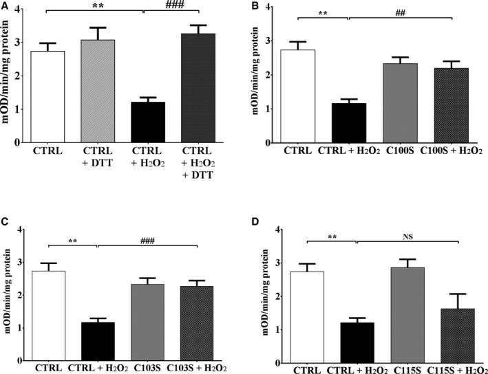
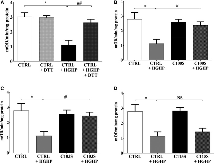
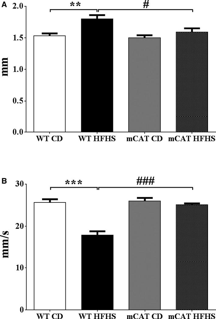
Methods and results: Mice fed a high-fat high-sucrose (HFHS) diet develop MHD with cardiac diastolic and mitochondrial dysfunction that is associated with oxidative posttranslational modifications of cardiac mitochondrial proteins. Transgenic mice that express catalase in mitochondria and wild-type mice were fed an HFHS or control diet for 4 months. Cardiac mitochondria from HFHS-fed wild-type mice had a 3-fold greater rate of H2O2 production (P=0.001 versus control diet fed), a 30% decrease in complex II substrate-driven oxygen consumption (P=0.006), 21% to 23% decreases in complex I and II substrate-driven ATP synthesis (P=0.01), and a 62% decrease in complex II activity (P=0.002). In transgenic mice that express catalase in mitochondria, all HFHS diet-induced mitochondrial abnormalities were ameliorated, as were left ventricular hypertrophy and diastolic dysfunction. In HFHS-fed wild-type mice complex II substrate-driven ATP synthesis and activity were restored ex vivo by dithiothreitol (5 mmol/L), suggesting a role for reversible cysteine oxidative posttranslational modifications. In vitro site-directed mutation of complex II subunit B Cys100 or Cys103 to redox-insensitive serines prevented complex II dysfunction induced by ROS or high glucose/high palmitate in the medium.

Conclusion: Mitochondrial ROS are pathogenic in MHD and contribute to mitochondrial dysfunction, at least in part, by causing oxidative posttranslational modifications of complex I and II proteins including reversible oxidative posttranslational modifications of complex II subunit B Cys100 and Cys103.

Keywords: metabolic heart disease; mitochondria; obesity; oxidative protein modifications; oxidative stress.


Figures

Figure 1
Figure 1
Increased cardiac mitochondrial H2O2 production in high‐fat high‐sucrose (HFHS) diet–fed mice is prevented by transgenic expression of mitochondrial catalase (mCAT). The mitochondrial H2O2 production rate is increased in cardiac mitochondria from wild‐type (WT) but not mCAT mice. A, H2O2 production rate with a complex I substrate (5 mmol/L pyruvate+5 mmol/L malate); (B) H2O2 production rate with a complex II substrate (5 mmol/L succinate) and inhibition of reverse electron transport (2 μmol/L rotenone). Values are mean±SEM; n=4 to 5; **P<0.01 vs WT control diet (CD); ***P<0.001 vs WT CD; ## P<0.01 vs WT HFHS; ### P<0.001 vs WT HFHS.
Figure 2
Figure 2
Decreases in complex I and II substrate–driven cardiac mitochondrial ATP synthesis rates in high‐fat high‐sucrose (HFHS) diet–fed mice are prevented in mitochondrial catalase (mCAT) mice. Cardiac mitochondrial complex I and II substrate–driven ATP synthesis rates are decreased in HFHS‐fed wild‐type (WT) but not mCAT mice. A, Complex I substrate–driven ATP synthesis rate (5 mmol/L pyruvate+5 mmol/L malate). B, Complex II substrate–driven ATP synthesis rate (5 mmol/L succinate+2 μmol/L rotenone). Values are mean±SEM; n=4 to 6; **P<0.01 vs WT control diet (CD); ***P<0.001 vs WT CD; ## P<0.01 vs WT HFHS.
Figure 3
Figure 3
Decreased cardiac mitochondrial maximal and uncoupled oxygen consumption in high‐fat high‐sucrose (HFHS) diet–fed mice is normalized in mitochondrial catalase (mCAT) mice. A, Complex I substrate–driven maximal oxygen consumption rate (state III). B, Complex I substrate–driven uncoupled (oligomycin 2 μmol/L) oxygen consumption rate (state IV). C, Complex II substrate–driven maximal oxygen consumption rate (state III). D, Complex II substrate–driven uncoupled (oligomycin 2 μmol/L) oxygen consumption rate (state IV). Values are mean±SEM; n=4 to 6; *P<0.05 vs wild‐type (WT) control diet (CD); **P<0.01 vs WT CD; # P<0.05 vs WT HFHS.
Figure 4
Figure 4
Decreased cardiac mitochondrial complex II activity in high‐fat high‐sucrose (HFHS) diet–fed mice is corrected by exposure to dithiothreitol (DTT; 5 mmol/L) ex vivo and prevented by overexpression of mitochondrial catalase (mCAT) in vivo. A, In wild‐type (WT) mice, complex II activity assessed by the reduction of ubiquinone to ubiquinol is decreased by HFHS feeding and restored by exposure to DTT (5 mmol/L) ex vivo. B, In mCAT mice, the HFHS‐induced decrease in complex II activity is prevented. C, In mCAT mice, DTT exposure ex vivo has no effect irrespective of diet. Values are mean±SEM; n=4 to 5; ***P<0.001 vs WT control diet (CD); ### P<0.001 vs WT HFHS.
Figure 5
Figure 5
Reversible cysteine oxidative posttranslational modifications (OPTMs) of cardiac mitochondrial complex II subunit B (succinate dehydrogenase B; SDHB) are increased in high‐fat high‐sucrose (HFHS) diet–fed wild‐type (WT) mice and prevented in mitochondrial catalase (mCAT) mice. A biotin switch assay was used to detect reversibly oxidized cysteines on SDHB. A, Representative blot in which “I” represents protein labeled by the antibody for SDHB and “Ox” represents the portion of total protein containing a reversible cysteine OPTM. B, Bar graph showing the mean values for the ratio of oxidized (Ox) to total (I+Ox) SDHB, reflecting the proportion of reversibly oxidized cysteines. MW, molecular weight. Values are mean±SEM; n=4 to 5; ***P<0.001 vs WT control diet (CD); ### P<0.001 vs WT HFHS.
Figure 6
Figure 6
Oxidation of succinate dehydrogenase B (SDHB) Cys100 and Cys103 inhibits mitochondrial complex II activity. Exposure of cells to H2O2 (500 μmol/L, 10 minutes) decreased complex II activity, measured as per Figure 4, in HEK 293T cells. SDHB in which Cys100Ser (C100S), Cys103Ser (C103S), or Cys115Ser (C115S) was mutated to a redox‐insensitive serine was expressed in HEK 293T cells, and complex II activity was measured after exposure to H2O2. A, H2O2 decreased complex II activity in HEK cells transfected with empty vector (CTRL), and activity was restored after incubation of cell lysate with dithiothreitol (DTT; 5 mmol/L). B, Expression of the SDHB C100S mutant prevents the H2O2‐mediated decrease in complex II activity. C, Expression of the SDHB C103S mutant prevents the H2O2‐mediated decrease in complex II activity. D, Expression of the SDHB C115S mutant has no effect on the H2O2‐mediated decrease in complex II activity. Values are mean±SEM; n=5; **P<0.01 vs CTRL; ## P<0.01 vs CTRL+H2O2; ### P<0.001 vs CTRL+H2O2.
Figure 7
Figure 7
Oxidation of succinate dehydrogenase B (SDHB) Cys100 and Cys103 inhibits mitochondrial complex II activity. Exposure of cells to high‐glucose/high‐palmitate (HGHP) media (14 hours) decreased complex II activity, measured as per Figure 4, in HEK 293T cells. SDHB in which Cys100Ser (C100S), Cys103Ser (C103S), or Cys115Ser (C115S) was mutated to a redox‐insensitive serine was expressed in HEK 293T cells, and complex II activity was measured after exposure to HGHP media. A, HGHP treatment decreased complex II activity in HEK cells transfected with empty vector (CTRL), and activity was restored after incubation of cell lysate with dithiothreitol (DTT; 5 mmol/L). B, Expression of the SDHB C100S mutant prevents the HGHP‐mediated decrease in complex II activity. C, Expression of the SDHB C103S mutant prevents the HGHP‐mediated decrease in complex II activity. D, Expression of the SDHB C115S mutant has no effect on the HGHP‐mediated decrease in complex II activity. Values are mean±SEM; n=3 to 4; *P<0.05 vs CTRL; # P<0.05 vs CTRL+HGHP; ## P<0.01 vs CTRL+HGHP.
Figure 8
Figure 8
Left ventricular hypertrophy and diastolic dysfunction induced by high‐fat high‐sucrose (HFHS) diet feeding for 4 months is prevented in mitochondrial catalase (mCAT) mice. A, Total wall thickness. B, Myocardial peak early diastolic velocity (Em). Values are mean±SEM; n=4 to 5; **P<0.01 vs wild‐type (WT) control diet (CD); ***P<0.001 vs WT CD; # P<0.05 vs WT HFHS; ### P<0.001 vs WT HFHS.
Figure 9
Figure 9
Schematic overview of the adverse effects of a high‐fat high‐sucrose (HFHS) diet on mitochondrial and cardiac function. HFHS diet feeding was associated with impaired ATP production and increased reactive oxygen species (ROS) generation in cardiac mitochondria. Decreased ATP production may contribute to diastolic dysfunction by limiting the function of highly ATP‐dependent enzymes such as sarcoplasmic reticulum Ca2+ ATPase (SERCA) that are needed for normal diastolic calcium homeostasis. ROS may also trigger multiple extramitochondrial signaling cascades involved in myocyte hypertrophy and the regulation of diastolic function.

References

    1. Heidenreich PA, Albert NM, Allen LA, Bluemke DA, Butler J, Fonarow GC, Ikonomidis JS, Khavjou O, Konstam MA, Maddox TM, Nichol G, Pham M, Pina IL, Trogdon JG; American Heart Association Advocacy Coordinating C, Council on Arteriosclerosis T, Vascular B, Council on Cardiovascular R, Intervention, Council on Clinical C, Council on E, Prevention, Stroke C . Forecasting the impact of heart failure in the United States: a policy statement from the American Heart Association. Circ Heart Fail. 2013;6:606–619. - PMC - PubMed
    1. Main ML, Rao SC, O'Keefe JH. Trends in obesity and extreme obesity among US adults. JAMA. 2010;303:1695; author reply 1695‐1696. - PubMed
    1. Thomas DM, Weedermann M, Fuemmeler BF, Martin CK, Dhurandhar NV, Bredlau C, Heymsfield SB, Ravussin E, Bouchard C. Dynamic model predicting overweight, obesity, and extreme obesity prevalence trends. Obesity (Silver Spring). 2014;22:590–597. - PMC - PubMed
    1. Ayalon N, Gopal DM, Mooney DM, Simonetti JS, Grossman JR, Dwivedi A, Donohue C, Perez AJ, Downing J, Gokce N, Miller EJ, Liang CS, Apovian CM, Colucci WS, Ho JE. Preclinical left ventricular diastolic dysfunction in metabolic syndrome. Am J Cardiol. 2014;114:838–842. - PMC - PubMed
    1. Bugger H, Abel ED. Molecular mechanisms of diabetic cardiomyopathy. Diabetologia. 2014;57:660–671. - PMC - PubMed

Publication types

MeSH terms

LinkOut - more resources