. 2016 Jan 26;113(4):1074-9.
doi: 10.1073/pnas.1520398113. Epub 2016 Jan 11.

Purinergic receptor P2RY12-dependent microglial closure of the injured blood-brain barrier

Affiliations

Purinergic receptor P2RY12-dependent microglial closure of the injured blood-brain barrier

Nanhong Lou et al. Proc Natl Acad Sci U S A. .

Abstract

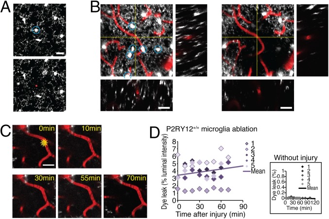
Microglia are integral functional elements of the central nervous system, but the contribution of these cells to the structural integrity of the neurovascular unit has not hitherto been assessed. We show here that following blood-brain barrier (BBB) breakdown, P2RY12 (purinergic receptor P2Y, G-protein coupled, 12)-mediated chemotaxis of microglia processes is required for the rapid closure of the BBB. Mice treated with the P2RY12 inhibitor clopidogrel, as well as those in which P2RY12 was genetically ablated, exhibited significantly diminished movement of juxtavascular microglial processes and failed to close laser-induced openings of the BBB. Thus, microglial cells play a previously unrecognized protective role in the maintenance of BBB integrity following cerebrovascular damage. Because clopidogrel antagonizes the platelet P2Y12 receptor, it is widely prescribed for patients with coronary artery and cerebrovascular disease. As such, these observations suggest the need for caution in the postincident continuation of P2RY12-targeted platelet inhibition.

Keywords: blood–brain barrier; clopidogrel; microglia; purinergic receptors; stroke.


Conflict of interest statement

The authors declare no conflict of interest.

Figures

Fig. 1.
Fig. 1.
Movement of juxtavascular microglia processes toward injured vessels requires P2RY12. (A) Representative time-lapse imaging of laser injury targeted outside the vasculature in CX3CR1+/EGFP/P2RY12+/+ mice (Upper), CX3CR1+/EGFP/P2RY12−/− mice (Middle), and CX3CR1+/EGFP/P2RY12+/+ mice receiving clopidogrel (Lower). (Scale bars, 25 µm.) (B, Upper) Juxtavascular microglial cell process accumulation in response to laser injury outside the vasculature as shown in A. Fluorescence signal of EGFP around the capillaries was normalized to fluorescence signal in the whole field. (Lower) Comparisons of the kinetics of process accumulation. n = 4–11 injuries from four animals; ns, P > 0.05; **P < 0.01, Kruskal–Wallis test. (C) Time-lapse images of juxtavascular microglial cell activation in response to laser injury in a CX3CR1+/EGFP/P2RY12+/+ mouse. The laser was targeted to the center of a capillary located 120 µm below the cortical surface (yellow star). Laser injury in a CX3CR1+/EGFP/P2RY12−/− mouse and a CX3CR1+/EGFP/P2RY12+/+ mouse treated with clopidogrel (20 mg/kg). (Scale bars, 20 µm.) (D, Upper) Kinetics of juxtavascular microglial process accumulation around the injured capillaries shown in C. Fluorescence signal of EGFP around the capillaries was normalized to fluorescence signal in the whole field. (Lower) Comparison of juxtavascular microglial cell processes around the injured capillary in CX3CR1+/EGFP/P2RY12+/+, CX3CR1+/EGFP/P2RY12−/−, and CX3CR1+/EGFP/P2RY12+/+ mice treated with clopidogrel; in CX3CR1+/EGFP/P2RY12+/+ mice treated with acetylsalicylic acid (10 mg/kg i.p.); and in CX3CR1+/EGFP/P2RY12+/+ mice treated with heparin (200 IU/kg i.v.). n = 5–9 capillaries from four to eight animals; ns, P > 0.05; *P < 0.05, **P < 0.01, one-way ANOVA with Tukey–Kramer test. (E, Upper) Tail bleeding time in vehicle control (n = 7), clopidogrel (5, 20, 30, 40, and 100 mg/kg i.p. daily for 3 d; n = 7–9), and acetylsalicylic acid (10 mg/kg, i.p. daily for 3 d, n = 5). (Lower) Platelet aggregation in whole blood from animals treated with vehicle control (n = 9–15), clopidogrel (5, 20, 30, 40, and 100 mg/kg i.p. daily for 3 d; n = 8–18), and acetylsalicylic acid (10 mg/kg, i.p. daily for 3 d; n = 11). (F) P2RY12 is predominantly, if not exclusively, expressed by juxtavascular microglial cells. DAPI (blue), CX3CR1-EGFP (green), P2RY12 (red), Laminin (white). Orthogonal views of XYZ stacked images are shown with planes of sections shown by white dotted lines. (Scale bar, 25 µm.) (G) Laser injury to a capillary did not induce accumulation of platelets at the injury site. Platelets were labeled with Calcein AM (green) inside capillaries labeled with Texas Red-dextran (red). Systemic administration of collagen (1 mg/mL) caused aggregations of platelets in random positions. (Scale bars, 20 µm.) (H, Upper) Kinetics of platelet accumulation inside the injured capillaries shown in G. (Lower) Comparison of platelet accumulation inside the capillary at 10 min after with or without the laser injury or collagen administration. n = 11 capillaries from four animals; ns, P > 0.05; **P < 0.01; one-way ANOVA with Tukey–Kramer test.
Fig. 2.
Fig. 2.
P2RY12 is required for rapid closure of the BBB, and juxtavascular microglia processes may temporarily seal BBB openings. (A) Experimental setup. The large MW weight tracer, Texas Red-dextran, was injected i.v. to outline the vasculature, and the small molecular Alexa Fluor 488 (10 µL, 80 μM) was repeatedly delivered by a catheter inserted into the internal carotid artery every 10 min to map the duration of closure of BBB openings induced by laser injury of single capillaries. (B, Upper panels) Time lapse of Alexa Fluor 488 (green) passage through a control, noninjured capillary. (Lower panels) Similar time lapse of Alexa Fluor 488 (green) passage through a capillary exposed to laser injury. The capillary is outlined by Texas Red-dextran (red). The dotted white square indicates the region used for quantification of Alexa Fluor 488 leakage. Alexa Fluor 488 leakage was defined as “peak fluorescence signal intensity outside the vessel divided by fluorescence signal intensity inside the vessel.” (Scale bar, 10 µm.) (C) Scatter histograms of Alexa Fluor 488 leakage in P2RY12+/+ mice (black), P2RY12−/−mice (orange), P2RY12+/+ mice receiving clopidogrel (20 mg/kg, red), acetylsalicylic acid (10 mg/kg, blue), or heparin (200 IU/kg, turquoise). Different color gradients indicate an individual set of capillaries. The lines indicate the average of linear regression curves, obtained by averaging the slopes and Y-intercept of each regression line from a single capillary. The average regression lines were used to obtain BBB closure time (X-intercept). (D) Summary histogram of BBB closure time. n = 4–7 capillaries from four to seven animals; ns, P > 0.05; **P < 0.01; one-way ANOVA with Tukey–Kramer test.
Fig. 3.
Fig. 3.
Laser injury induces accumulatation of juxtavascular microglia processes and does not affect capillary perfusion. (A) Electron microscopic image of laser injury in cerebral cortex. Yellow dotted line with a yellow star indicates the site of the focal injury. Green dotted line indicates the accumulated juxtavascular microglia processes, with arrows indicating the close apposition of adjacent microglia processes extended toward the injury site. (B) Immunohistochemical analysis of focal laser injury site in cerebral cortex of CX3CR1+/EGFP animals. P2RY12 and E-cadherin (red) colocalized with EGFP (green) and are highly expressed in microglial cell processes encircling the injury site. In contrast, occludin (red) was detected only in vascular endothelial cells, but not in microglial cells. (Scale bars, 20 µm.) (C) Time lapse of a laser-injured capillary (red) with microglia (gray) in CX3CR1+/EGFP/P2RY12+/+, CX3CR1+/EGFP/P2RY12−/−, and CX3CR1+/EGFP/P2RY12+/+ mice treated with 20 mg/kg clopidogrel. (Scale bars, 10 µm.) (D) Plots of capillary diameter at the site of laser injury plotted as a function of time in CX3CR1+/EGFP/P2RY12+/+, CX3CR1+/EGFP/P2RY12−/−, and CX3CR1+/EGFP/P2RY12+/+ mice treated with 20 mg/kg clopidogrel. n = 3–5 capillaries from three to five animals. (E, Left) Strategy for collecting time series of XT line-scan images in capillaries filled with Texas Red-dextran (red). (Right) Line scans were collected at 0–64 min after laser injury. (F) Plots of RBC velocity and flux of capillary exposed to laser injury in P2RY12+/+, P2RY12−/−, and P2RY12+/+ mice treated with 20 mg/kg clopidogrel. n = 5–12 capillaries from three animals.
Fig. 4.
Fig. 4.
Juxtavascular microglia ablation attenuates vascular closure. (A) Ablation of juxtavascular microglial cell. Propidium iodide (30 µM) was applied after a juxtavascular microglia was ablated with focused laser radiation. Only the ablated microglial cell (blue circle) that lost EGFP fluorescence was stained with a cell death marker propidium iodide (red). (Scale bar, 20 µm.) (B) Projection images (55 µm in z direction) and orthogonal views (XZ and YZ planes at yellow dotted lines) of a field before (Left) and after (Right) laser ablation of six juxtavascular microglial cells (white with blue circles) located within a radius of 40 µm around the target capillary (red, at the crosshair) in a CX3CR1+/EGFP/P2RY12+/+ mouse. (Scale bars, 20 µm.) (C) Time series of experiment with ablation of juxtavascular microglial cells shows that the region around the injured capillary remained free of microglial cell processes for the duration of the experiment. No juxtavascular microglial cell processes were in contact with the injured capillary at 70 min. (Scale bar, 20 µm.) (D) Scatter histogram of Alexa Fluor 488 leakage in microglia-ablated CX3CR1+/EGFP/P2RY12+/+ mice. Different color gradients indicate an individual set of capillaries. The line indicates the average of linear regression curves (y = 0.0144x–1125.84), obtained by averaging slopes and Y-intercept of each regression line from each capillary. Rate of BBB closure was 1.44 ± 0.87%/min (n = 5 capillaries), indicating that the leak worsened over time rather than gradually closing. (Inset) Scatter histogram of Alexa Fluor 488 leakage following juxtavascular microglial cell ablation but without laser injury to the capillary. The line indicates the average of linear regression curves (y = 0.00021x–0.01092; n = 5 capillaries).

References

    1. Aguzzi A, Barres BA, Bennett ML. Microglia: Scapegoat, saboteur, or something else? Science. 2013;339(6116):156–161. - PMC - PubMed
    1. Marín-Teva JL, et al. Microglia promote the death of developing Purkinje cells. Neuron. 2004;41(4):535–547. - PubMed
    1. Paolicelli RC, et al. Synaptic pruning by microglia is necessary for normal brain development. Science. 2011;333(6048):1456–1458. - PubMed
    1. Schafer DP, et al. Microglia sculpt postnatal neural circuits in an activity and complement-dependent manner. Neuron. 2012;74(4):691–705. - PMC - PubMed
    1. Tremblay ME, Lowery RL, Majewska AK. Microglial interactions with synapses are modulated by visual experience. PLoS Biol. 2010;8(11):e1000527. - PMC - PubMed

Publication types

LinkOut - more resources