Comparative Study
. 2016 Oct;100(10):1377-82.
doi: 10.1136/bjophthalmol-2015-307480. Epub 2016 Jan 11.

Wide field of view swept-source optical coherence tomography for peripheral retinal disease

Affiliations
Comparative Study

Wide field of view swept-source optical coherence tomography for peripheral retinal disease

Ryan P McNabb et al. Br J Ophthalmol. 2016 Oct.

Abstract

Background/aims: To assess peripheral retinal lesions and the posterior pole in single widefield optical coherence tomography (OCT) volumes.

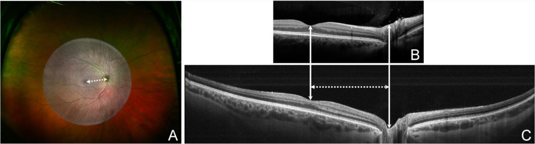
Methods: A wide field of view (FOV) swept-source OCT (WFOV SSOCT) system was developed using a commercial swept-source laser and a custom sample arm consisting of two indirect ophthalmic lenses. Twenty-seven subjects with peripheral lesions (choroidal melanomas, choroidal naevi, sclerochoroidal calcification, retinitis pigmentosa, diabetic retinopathy, retinoschisis and uveitis) were imaged with the WFOV SSOCT. Volumes were taken in primary gaze. Using the optic nerve to fovea distance as a reference measurement, comparisons were made between the lateral FOV of the WFOV SSOCT, current generation spectral-domain OCT (SDOCT) and widefield scanning laser ophthalmoscopy (SLO) of the same eyes.

Results: Peripheral pathologies were captured with WFOV SSOCT in 26 of the 27 subjects. The one not captured was in the far nasal periphery and was not seen in the primary gaze volume. Posterior pole associated pathologies were captured in all subjects. Current generation SDOCT had a mean lateral FOV of 2.08±0.21 optic nerve to fovea distance units, WFOV SSOCT had an FOV of 4.62±0.62 units and SLO had an FOV of 9.35±1.02 units.

Conclusions: WFOV OCT can be used to examine both peripheral retinal pathology and the posterior pole within a single volume acquisition. SLO had the greatest FOV, but does not provide depth information. Future studies using widefield OCT systems will help further delineate the role of WFOV OCT to quantitatively assess and monitor peripheral retinal disease in three dimensions.

Keywords: Imaging; Retina.


Figures

Figure 1
Figure 1. Field of view (FOV) measurement in a normal subject for A. wide field scanning laser ophthalmoscope (SLO), B. current generation spectral domain optical coherence tomography (SDOCT, Heidelberg), and C. wide FOV swept source OCT (SSOCT) in a normal subject
The greyscale summed voxel projection from the wide FOV SSOCT volume is overlaid over the SLO image in 1A for comparison. The white dotted line represents the optic nerve to fovea distance and is used as the reference unit of measurement for the horizontal FOV of each system. In this subject, the current generation SDOCT has a horizontal FOV of 1.9 optic nerve-to-fovea distance units, the wide FOV SSOCT has a FOV of 4.2 units, and the wide-field SLO has a FOV of 9.3 units.
Figure 2
Figure 2. Wide FOV images of a choroidal nevus (white arrows) and associated subretinal fluid
A: Red/green and B: autofluorescence SLO images. C: Wide FOV swept source OCT (WFOV SSOCT) summed voxel projection composed of B scans (blue and green lines indicate cross sectional scans in 2D and 2E). Subtle elevation of the retinal vessels over the nevus can be seen. D: WFOV SSOCT averaged B scan through fovea and optic nerve head demonstrating normal foveal contour with preserved external limiting membrane and ellipsoid zone. E: Unaveraged B scan from same WFOV SSOCT volume as Top Middle image at a custom angle showing the peripheral choroidal nevus with subretinal fluid at the nasal margin of the nevus, mild overlying retinal pigment epithelium alteration, absence of overlying drusen, and loss of choriocapillaris over the lesion. The posterior margin of the nevus was not completely visualized. The graininess in the images is due to the unaveraged nature of this arbitrarily cut B scan.
Figure 3
Figure 3. Wide FOV images of a calcified scleral mass seen in the superotemporal macular region (white arrow)
A: Red/green SLO. B: Standard field of view clinical SDOCT B-scan showing only the lesion due to limits on field of view. Scleral thickening compressing the overlying choroid is seen. C: Three-dimensional rendering of wide field of view swept source OCT (WFOV SSOCT) volume with large elevated mass (white arrow), fovea (green arrow), and optic nerve head (blue arrow). Fine horizontal “ripples” on the retinal surface are due to patient motion captured during the volume scan. D: Un-averaged custom oriented WFOV SSOCT B scan through data in 3C showing both the lesion and optic nerve head in the same scan. E: Averaged horizontal WFOV SSOCT B scan through lesion. In both 3D and 3E, scleral thickening with overlying choroidal atrophy due to compression with loss of choriocapillaris and retinal pigment epithelial atrophy is seen. There is no subretinal fluid present.
Figure 4
Figure 4. Wide FOV images of retinitis pigmentosa (RP) and a lamellar macular hole
A: Red/green SLO image. B: Wide FOV swept source OCT (WFOV SSOCT) summed volume projection with blue arrow locating the C: WFOV SSOCT arbitrary direction B-scan through fovea and optic nerve head. The foveal area has a lamellar macular hole (arrowhead), extensive outer retinal atrophy with loss of subfoveal external limiting membrane and ellipsoid zone, and alteration of subfoveal choroidal morphology. Peripherally in the same B-scan, advanced retinitis pigmentosa with significant choroidal and retinal atrophy temporally and nasally with inner retinal hyperreflectivity corresponding to the bone spicules is present. The graininess in the image is due to the unaveraged nature of this arbitrarily cut B scan.
Figure 5
Figure 5. Wide FOV OCT images of proliferative diabetic retinopathy (PDR) with retinochisis, PDR with peripheral ischemia, and also images of uveitis
PDR with retinochisis. A: Wide FOV swept source OCT (WFOV SSOCT) summed volume projection with blue arrow locating the cross sectional scan in B and the green arrow the cross sectional scan in C. B: WFOV SSOCT B-scan through fovea and optic nerve head. The temporal retina shows retinoschisis, and there is absence of macular edema (white arrow) with relative preservation of the outer retinal bands. C: WFOV SSOCT B-scan through the area of peripheral retinoschisis showing the area of vitreous traction corresponding to the inactive fibroavascular proliferation. WFOV SSOCT allowed confirmation of absence of inner retinal breaks in the areas of retinoschisis. PDR with peripheral ischemia. D: Wide field SLO fluorescein angiogram (late phase) showing a preserved central macular island of perfusion with extensive peripheral ischemia and staining corresponding to dense pan retinal photocoagulation scars. E: WFOV SSOCT summed volume projection with blue arrow locating the cross section in F and the green arrow the cross sectional scan in G. F: WFOV SSOCT B-scan through fovea and optic nerve head. There is disorganization of the retinal inner layers in the macula, absence of diabetic macular edema and severe peripheral retinal thinning and atrophy (also in G) corresponding to the peripheral ischemia seen on the fluorescein angiogram. Uveitis (Vogt-Koyanagi-Harada syndrome). H: Red/green SLO image with macular SDOCT inset (arrow indicates approximate scale) taken at initial presentation. There is a pronounced serous retinal detachment involving the macula and extending nasally. The SDOCT obtained at this initial presentation showed subretinal fluid with hyperreflective material extending beyond the margins of the scan and loss of outer retinal layers and granular deposits anterior to the retinal pigment epithelium. I: WFOV SSOCT summed volume projection with blue arrow locating the cross section in J and the green arrow locating the cross section in K. J: WFOV SSOCT scan through the fovea obtained 2 months after starting immunosuppressive therapy showing significant improvement in the subretinal fluid with persistent mild choroidal thickening. There is a thin residual sliver of subfoveal subretinal fluid (inset) with subretinal hyperreflective dots. K: WFOV SSOCT B-scan through a different area of the volume. Residual subretinal fluid with hyperreflective dots is also present here with loss of outer retinal structures. The yellow arrow corresponds to the location in the projection in I. Other localized areas similar to this were present throughout this volume.

References

    1. Huang D, Swanson EA, Lin CP, Schuman JS, Stinson WG, Chang W, Hee MR, Flotte T, Gregory K, Puliafito CA, et al. Optical coherence tomography. Science. 1991 Nov 22;254(5035):1178–1181. - PMC - PubMed
    1. Swanson EA. OCT technology transfer and the OCT market. In: Drexler W, Fujimoto JG, editors. Optical coherence tomography. New York: Springer; 2015. pp. 2529–2571.
    1. Schuman JS, Puliafito CA, Fujimoto JG, Duker JS. Optical coherence tomography of ocular disease. 3rd. Thorofare, NJ: SLACK incorporated; 2012. pp. 3–26.
    1. Shields CL, Materin MA, Shields JA. Review of optical coherence tomography for intraocular tumors. Curr Opin Ophthalmol. 2005 Jun;16(3):141–154. - PubMed
    1. Torres VL, Brugnoni N, Kaiser PK, et al. Optical coherence tomography enhanced depth imaging of choroidal tumors. Am J Ophthalmol. 2011 Apr;151(4):586–593. - PubMed

Publication types