. 2016 Mar 31;127(13):1719-27.
doi: 10.1182/blood-2015-10-676114. Epub 2016 Jan 11.

What is going on between defibrotide and endothelial cells? Snapshots reveal the hot spots of their romance

Affiliations

What is going on between defibrotide and endothelial cells? Snapshots reveal the hot spots of their romance

Marta Palomo et al. Blood. .

Abstract

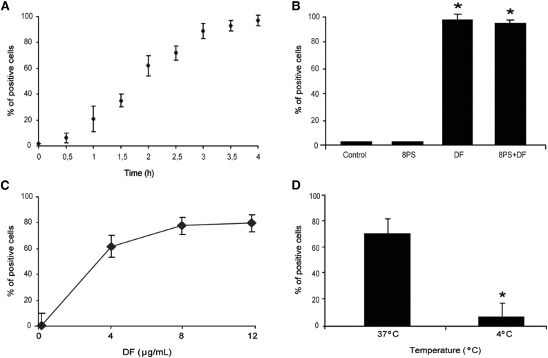
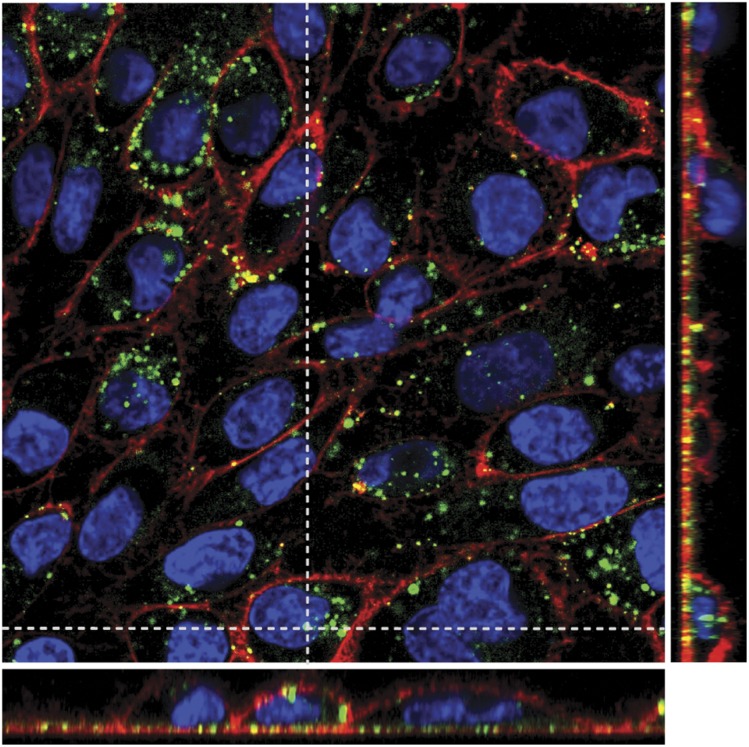
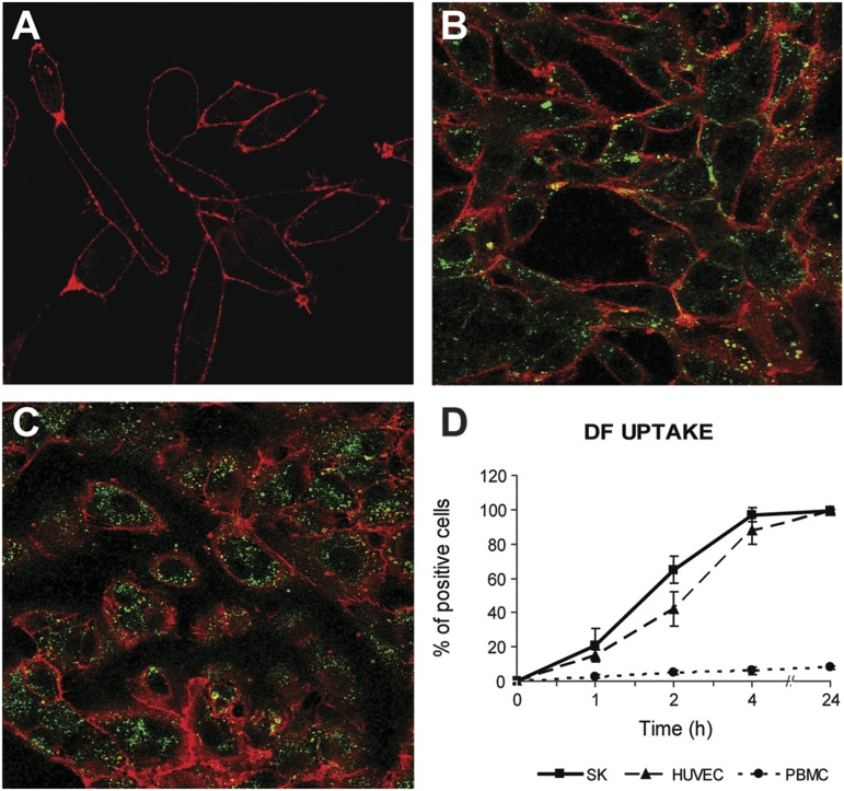
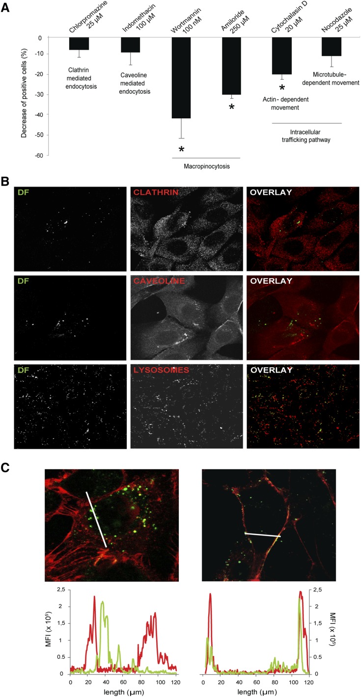
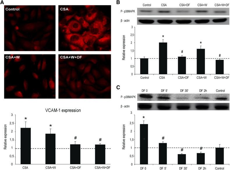
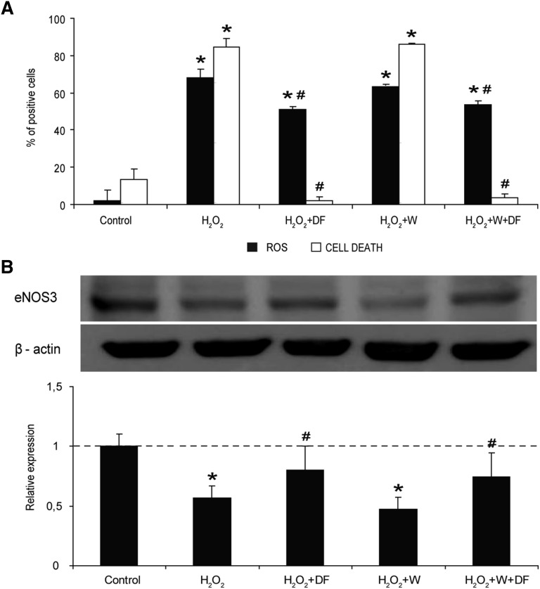
Defibrotide (DF) has received European Medicines Agency authorization to treat sinusoidal obstruction syndrome, an early complication after hematopoietic cell transplantation. DF has a recognized role as an endothelial protective agent, although its precise mechanism of action remains to be elucidated. The aim of the present study was to investigate the interaction of DF with endothelial cells (ECs). A human hepatic EC line was exposed to different DF concentrations, previously labeled. Using inhibitory assays and flow cytometry techniques along with confocal microscopy, we explored: DF-EC interaction, endocytic pathways, and internalization kinetics. Moreover, we evaluated the potential role of adenosine receptors in DF-EC interaction and if DF effects on endothelium were dependent of its internalization. Confocal microscopy showed interaction of DF with EC membranes followed by internalization, though DF did not reach the cell nucleus even after 24 hours. Flow cytometry revealed concentration, temperature, and time dependent uptake of DF in 2 EC models but not in other cell types. Moreover, inhibitory assays indicated that entrance of DF into ECs occurs primarily through macropinocytosis. Our experimental approach did not show any evidence of the involvement of adenosine receptors in DF-EC interaction. The antiinflammatory and antioxidant properties of DF seem to be caused by the interaction of the drug with the cell membrane. Our findings contribute to a better understanding of the precise mechanisms of action of DF as a therapeutic and potential preventive agent on the endothelial damage underlying different pathologic situations.


Figures

Figure 1
Figure 1
Uptake of defibrotide by endothelial cells. (A) SK cells were incubated with DF (4 μg/mL final concentration), labeled with Alexa 488, for the indicated period of time (from 0-4 h). (B) The addition of 10 μM adenosine receptor antagonist (8-p-sulfophenyltheophylline) to SK monolayers for 1 hour before the incubation with DF (4 h) did not interfere with SK–DF interaction. (C) For saturation assays, SK monolayers were exposed to increasing doses of DF (from 0-12 μg/mL) for 2 hours. (D) For temperature dependence assays, SK were incubated with 4 μg/mL for 2 hours at 37°C and 4°C. Results are expressed in percentage of positive cells.
Figure 2
Figure 2
DF internalization by SK cells. Merged confocal images of all individual channels (red staining with wheat germ agglutinin for membranes, blue with Hoechst for nuclei, and green with Alexa 488 for DF), and Z projection (narrow images in the right and in the bottom of the main picture) show that DF remains in the cytoplasm displaying vesicular staining and does not enter into the nuclei, at least after 24 hours of incubation. Confocal images were taken using a Leica TCS SP5 microscope and a 63× oil immersion objective. Optical sections (z) were performed each 2 μm. Image analysis was performed using Fiji software (National Institutes of Health).
Figure 3
Figure 3
Specificity of DF labeling and DF interaction with ECs. (A) Confocal microscopy images show no green staining when SK cells (red staining with wheat germ agglutinin for membranes) were incubated only with Alexa 488 compared with (B) the incubation of SK cells with DF labeled with Alexa 488. (C) Exposure of HUVEC to DF (4 μg/mL) for 4 hours also resulted in the internalization of the drug. (D) Flow cytometry results show that SK and HUVEC follow the same DF interaction kinetics. PBMCs do not interact with DF. Confocal images were taken using a Leica TCS SP5 microscope and a 63× oil immersion objective. Image analysis was performed using Fiji software (National Institutes of Health).
Figure 4
Figure 4
DF is internalized by ECs through macropinocytic mechanisms. (A) Bar diagram shows the decrease in the uptake of DF by SK cells in the presence of endocytosis and vesicle-trafficking inhibitors. Data obtained from flow cytometry experiments are expressed as mean ± standard error of the mean, n = 6, being *P < .05 vs 100% of positive cells for DF in the absence of the inhibitors. (B) Confocal microscopy images correspond to the negative results of colocalization assays between DF (green, labeled with Alexa 488) and clathrin, caveoline, and lysosomes (first, second, and third lines, respectively). SK cells were incubated with DF for 15 minutes to evaluate colocalization with clathrin and caveoline, and for 6 hours to evaluate DF–lysosomal interaction. (C) Images to the left and right correspond to SK cells incubated with DF (green) in the absence or presence of Wortmannin (W), respectively (red staining with wheat germ agglutinin for membranes). Graphs above represent mean fluorescence intensity and follow the same distribution. DF staining inside the cells can be visualized in the absence of W (left image, left graphic). DF staining is attached to the membrane in the presence of W (right image, right graphic). Confocal images were taken using a Leica TCS SP5 microscope and a 63× oil immersion objective. Image analysis was performed using Fiji software (National Institutes of Health).
Figure 5
Figure 5
Inflammation of ECs is prevented by DF interaction with the cell membrane. (A) Micrographs show VCAM-1 expression on SK exposed to CSA (200 ng/mL, 24 h), without and with DF in the media and in the absence and presence of Wortmannin (W), as indicated. Bar diagrams represent levels of VCAM-1 expression on SK. (B) Activation of p38 MAPK in SK cells by CSA, without and with DF and in the absence and presence of W. Immunoblot image shows phosphorylated p38 MAPK, and the bar diagram represents the relative quantification (vs control). (C) Activation of p38 MAPK by CSA in SK cells previously incubated with DF for different time points. The immunoblot image shows phosphorylated p38 MAPK and the bar diagram represents the relative quantification (vs control). The dotted line represents the mean expression in control cells. All data correspond to relative expression, n = 4, being *P < .05 vs control and #P < .05 vs CSA.
Figure 6
Figure 6
The antioxidant effect of DF on ECs is caused by its interaction with the cell membrane. (A) Flow cytometry experiments reveal that DF has an antioxidant effect in front of an oxidative stimuli (H2O2), even in the presence of Wortmannin (W). Black bars correspond to intracellular ROS in SK cells induced by incubation with 1 μM H2O2, expressed as percentage of positive cells. White bars correspond to percentage of dead cells after exposing SK to a high concentration of H2O2 (50 μM). (B) The immunoblot image shows changes in the presence of the protein eNOS3 in SK exposed to 1 μM H2O2, without and with DF in the media and in the absence and presence of Wortmannin (W), as indicated. The bar diagram represents relative presence of eNOS3 vs control. Data corresponds to n = 4, being *P < .05 vs control and #P < .05 vs H2O2.

Comment in

References

    1. Richardson PG, Corbacioglu S, Ho VT, et al. Drug safety evaluation of defibrotide. Expert Opin Drug Saf. 2013;12(1):123–136. - PubMed
    1. Espinosa G, Berman H, Cervera R. Management of refractory cases of catastrophic antiphospholipid syndrome. Autoimmun Rev. 2011;10(11):664–668. - PubMed
    1. Francischetti IM, Oliveira CJ, Ostera GR, et al. Defibrotide interferes with several steps of the coagulation-inflammation cycle and exhibits therapeutic potential to treat severe malaria. Arterioscler Thromb Vasc Biol. 2012;32(3):786–798. - PMC - PubMed
    1. Pescador R, Capuzzi L, Mantovani M, Fulgenzi A, Ferrero ME. Defibrotide: properties and clinical use of an old/new drug. Vascul Pharmacol. 2013;59(1-2):1–10. - PubMed
    1. Palomo M, Diaz-Ricart M, Carbo C, et al. The release of soluble factors contributing to endothelial activation and damage after hematopoietic stem cell transplantation is not limited to the allogeneic setting and involves several pathogenic mechanisms. Biol Blood Marrow Transplant. 2009;15(5):537–546. - PubMed

Publication types

MeSH terms