. 2016 Mar 11;291(11):5774-5787.
doi: 10.1074/jbc.M115.685321. Epub 2016 Jan 11.

Regulatory Characteristics of Vibrio vulnificus gbpA Gene Encoding a Mucin-binding Protein Essential for Pathogenesis

Affiliations

Regulatory Characteristics of Vibrio vulnificus gbpA Gene Encoding a Mucin-binding Protein Essential for Pathogenesis

Kyung Ku Jang et al. J Biol Chem. .

Abstract

Binding to mucin is the initial step for enteropathogens to establish pathogenesis. An open reading frame, gbpA, of Vibrio vulnificus was identified and characterized in this study. Compared with wild type, the gbpA mutant was impaired in binding to mucin-agar and the mucin-secreting HT29-methotrexate cells, and the impaired mucin binding was restored by the purified GbpA provided exogenously. The gbpA mutant had attenuated virulence and ability of intestinal colonization in a mouse model, indicating that GbpA is a mucin-binding protein and essential for pathogenesis of V. vulnificus. The gbpA transcription was growth phase-dependent, reaching a maximum during the exponential phase. The Fe-S cluster regulator (IscR) and the cyclic AMP receptor protein (CRP) coactivated, whereas SmcR, a LuxR homologue, repressed gbpA. The cellular levels of IscR, CRP, and SmcR were not significantly affected by one another, indicating that the regulator proteins function cooperatively to regulate gbpA rather than sequentially in a regulatory cascade. The regulatory proteins directly bind upstream of the gbpA promoter PgbpA. DNase I protection assays, together with the deletion analyses of PgbpA, demonstrated that IscR binds to two specific sequences centered at -164.5 and -106, and CRP and SmcR bind specifically to the sequences centered at -68 and -45, respectively. Furthermore, gbpA was induced by exposure to H2O2, and the induction appeared to be mediated by elevated intracellular levels of IscR. Consequently, the combined results indicated that IscR, CRP, and SmcR cooperate for precise regulation of gbpA during the V. vulnificus pathogenesis.

Keywords: CRP; GbpA; IscR; LuxR homologue; Vibrio vulnificus; bacterial adhesion; gene regulation; mucin; oxidative stress; quorum sensing.


Figures

FIGURE 1.
FIGURE 1.
Effect of GbpA on mucin binding and host cell adhesion of V. vulnificus. Upper panel, mucin binding activities of the strains. A, strains (∼107 cfu) were added to each well of 12-well culture dishes containing the mucin-agar and various amounts of GbpA provided exogenously as indicated. After 1 h of incubation, the adherent bacterial cells were enumerated as cfu per well. WT, wild type; gbpA, gbpA mutant. B, mucin-secreting HT29-MTX cells (∼107 cells) seeded in each well of 12-well culture dishes were infected at a multiplicity of infection of 10 with the strains as indicated. After a 30-min incubation, the adherent bacterial cells were enumerated as cfu per well. Error bars represent the S.D. *, p < 0.05, and **, p < 0.005 relative to the wild type. WT (pJK1113, empty vector), wild type; gbpA (pJK1113), gbpA mutant; gbpA (pKK1402), complemented strain. Lower panel, development of the mucin-secreting HT29-MTX cells. C, bright field image of HT29-MTX cells. D, nucleus of HT29-MTX cells was stained blue with DAPI. E, mucin of HT29-MTX cells was stained green with the anti-MUC5AC primary antibody and then labeled with FITC-conjugated secondary antibody. F, merged image of C–E. Images are visualized using a confocal laser scanning microscope. Scale bar, 40 μm.
FIGURE 2.
FIGURE 2.
Mouse lethality and colonization activity of the V. vulnificus strains. A, 7-week-old specific pathogen-free female ICR mice were intragastrically infected with the wild type (WT) or the gbpA mutant (gbpA) (Table 1) at doses of ∼109 cfu as indicated. Mouse survival was monitored for 24 h. B, four mice were intragastrically infected with an inoculum prepared by mixing equal numbers of MORR and KK141 (Table 1), and then the bacterial cells colonized on the small intestine were enumerated as cfu at the indicated time intervals. Each circle corresponds to the ratio of cfu recovered from the intestines to the cfu inoculated (the colonization index) for an individual mouse. The median values are displayed as a solid line (MORR) or dashed line (KK141) on the graph. WT, wild type; gbpA, gbpA mutant; MORR, wild type with rifampicin resistance; KK141, gbpA mutant with rifampicin and streptomycin resistance.
FIGURE 3.
FIGURE 3.
Effects of growth phases and global regulatory proteins on the gbpA expression. Upper panel, growth kinetics of V. vulnificus and growth phase-dependent expression of gbpA. A, growth of the wild type culture in LBS was monitored spectrophotometrically at 600 nm (A600), and total RNAs were isolated from the cells harvested at different growth phases (from left, at A600 of 0.5, 1.0, 1.5, 2.0, and 2.5) as indicated by arrows. B, gbpA mRNA levels were determined by qRT-PCR analyses, and the gbpA mRNA level in the cells grown to an A600 of 0.5 was set as 1. Error bars represent the S.D. *, p < 0.05, and **, p < 0.005 relative to the cells grown to an A600 of 0.5. Lower panel, expression of gbpA in V. vulnificus with different genetic backgrounds. Samples were harvested from the cultures of the wild type (WT) and isogenic mutants grown aerobically to an A600 of 0.5 and analyzed to determine the gbpA mRNA and GbpA protein levels. C and E, gbpA mRNA levels were determined by qRT-PCR analyses, and the gbpA mRNA level in the wild type was set as 1. **, p < 0.005 relative to the wild type. D and F, protein samples were resolved by SDS-PAGE, and GbpA was detected by Western blotting using the rabbit anti-V. vulnificus GbpA serum. (pJK1113), wild type; iscR (pJK1113), iscR mutant; smcR (pJK1113), smcR mutant; crp (pJK1113), crp mutant; iscR (pKK1403), crp (pKK1404), and smcR (pKK1405), complemented strains.
FIGURE 4.
FIGURE 4.
IscR and CRP coactivate gbpA additively. Samples were harvested from the cultures of the wild type (WT) and isogenic mutants grown aerobically to an A600 of 0.5 and analyzed to determine the gbpA mRNA levels and IscR and CRP protein levels. The gbpA mRNA levels were determined by qRT-PCR analyses, and the gbpA mRNA level in the wild type was set as 1. Error bars represent the S.D. The cellular levels of IscR and CRP were determined by Western blot analyses using the rabbit anti-V. vulnificus IscR and anti-V. vulnificus CRP sera, respectively. WT, wild type; iscR, iscR mutant; crp, crp mutant; iscR crp, iscR crp double mutant.
FIGURE 5.
FIGURE 5.
Cellular levels of IscR, CRP, and SmcR are unaffected by one another. The wild type and isogenic mutants were grown aerobically to an A600 of 0.5 (log phase, L) and 2.0 (stationary phase, S). The cells were then examined for the presence of IscR, CRP, SmcR, and GbpA proteins by Western blot analyses using the rabbit anti-V. vulnificus IscR, anti-V. vulnificus CRP, and anti-V. vulnificus SmcR, anti-V. vulnificus GbpA sera, respectively. WT, wild type; iscR, iscR mutant; crp, crp mutant; smcR, smcR mutant.
FIGURE 6.
FIGURE 6.
Transcription start site and sequences of the gbpA regulatory region. A, transcription start site of gbpA was determined by primer extension of the RNA isolated from the wild type grown aerobically to an A600 of 0.5. Lanes C, T, A, and G represent the nucleotide sequencing ladders of pKK1401. The asterisk indicates the transcription start site of gbpA. B, transcription start site of gbpA is indicated by a bent arrow, and the positions of the putative −10 and −35 regions are underlined. The sequences for binding of IscR (ISCRB1 and ISCRB2, white boxes), CRP (CRPB, shaded boxes), and SmcR (SMCRB, gray boxes) were determined later in this study (Fig. 10). The nucleotides showing enhanced cleavage are indicated by black boxes. The consensus sequences for binding of IscR (type 2), CRP, and SmcR are, respectively, indicated above the V. vulnificus DNA sequence. R, A or G; Y, C or T; W, A or T; x, any nucleotide.
FIGURE 7.
FIGURE 7.
Deletion analysis of the PgbpA regulatory region. A, construction of gbpA-lux fusion pKK reporters. PCR fragments carrying the gbpA regulatory region with 5′ end deletions were subcloned into pBBR-lux (44) to create each pKK reporter. Solid lines, the upstream region of gbpA; black blocks, the gbpA coding region; open blocks, luxCDABE. The wild type gbpA regulatory region is shown on top with the proposed −10 and −35 regions, and the binding sites for IscR (ISCRB1 and ISCRB2, white boxes), CRP (CRPB, shaded box), and SmcR (SMCRB, gray box) were determined later in this study (Fig. 10). B, cellular luminescence determined from the wild type (black bars), iscR mutant (dark gray bars), crp mutant (gray bars), and smcR mutant (open bars) containing each pKK reporter as indicated. Cultures grown aerobically to an A600 of 0.5 were used to measure the cellular luminescence. Error bars represent the S.D. WT, wild type; iscR, iscR mutant; crp, crp mutant; smcR, smcR mutant.
FIGURE 8.
FIGURE 8.
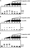
Specific bindings of IscR, CRP, and SmcR to PgbpA. A 390-bp DNA fragment of the gbpA regulatory region was radioactively labeled and then used as a probe DNA. The radiolabeled probe DNA (5 nm) was mixed with increasing amounts IscR (A), CRP (B), and SmcR (C) as indicated. For competition analysis, the same but unlabeled 390-bp DNA fragment was used as a self-competitor DNA. Various amounts of the self-competitor DNA were added to a reaction mixture containing the 5 nm labeled DNA prior to the addition of 30 nm IscR (A), 30 nm CRP (B), and 100 nm SmcR (C). B, bound DNA; F, free DNA.
FIGURE 9.
FIGURE 9.
Sequences for binding of IscR, CRP, and SmcR to PgbpA. A 390-bp DNA fragment of the gbpA regulatory region was radioactively labeled and then used as a DNA probe. The radiolabeled DNA probe (25 nm) was incubated with increasing amounts of IscR (A), CRP (B), and SmcR (C) as indicated. The regions protected by IscR, CRP, and SmcR are indicated by white boxes, shaded boxes, and gray boxes, respectively. The nucleotides showing enhanced cleavage are indicated by black boxes. Lanes C, T, A, and G represent the nucleotide sequencing ladders of pKK1401.
FIGURE 10.
FIGURE 10.
Effects of oxidative stress and apo-IscR on the activity of PgbpA. Total RNAs and proteins were isolated either from the cultures grown anaerobically to an A600 of 0.5 and then exposed to various concentrations of H2O2 for 10 min as indicated (A and B) or from the cultures grown aerobically to an A600 of 0.5 (C and D). A and C, gbpA mRNA levels were determined by qRT-PCR analyses, and the gbpA mRNA level in the wild type unexposed to H2O2 (A) or the wild type (C) was set to 1. Error bars represent the S.D. **, p < 0.005 relative to the wild type unexposed to H2O2 (A) or to the wild type (C). B and D, protein samples were resolved by SDS-PAGE, and IscR (or IscR3CA), CRP, SmcR, and GbpA were detected by Western blotting using the rabbit anti-V. vulnificus IscR, anti-V. vulnificus CRP, anti-V. vulnificus SmcR, and anti-V. vulnificus GbpA sera, respectively. WT, wild type; iscR, iscR mutant; iscR3CA, a strain expressing apo-locked IscR3CA.

Similar articles

Cited by

References

    1. McGuckin M. A., Lindén S. K., Sutton P., and Florin T. H. (2011) Mucin dynamics and enteric pathogens. Nat. Rev. Microbiol. 9, 265–278 - PubMed
    1. Neutra M. R., and Forstner J. F. (1987) in Physiology of the Gastrointestinal Tract (Johnson L. R., ed) Vol. 2, 2nd Ed., pp. 975–1009, Raven Press, Ltd., New York
    1. Wiggins R., Hicks S. J., Soothill P. W., Millar M. R., and Corfield A. P. (2001) Mucinases and sialidases: their role in the pathogenesis of sexually transmitted infections in the female genital tract. Sex Transm. Infect. 77, 402–408 - PMC - PubMed
    1. Thornton D. J., and Sheehan J. K. (2004) From mucins to mucus: toward a more coherent understanding of this essential barrier. Proc. Am. Thorac. Soc. 1, 54–61 - PubMed
    1. Ofek I., Bayer E. A., and Abraham S. N. (2013) in The Prokaryotes (Rosenberg E., Delong E. F., Lory S., Stackebrandt E., and Thompson F., eds) pp. 107–123, 10.1007/978-3-642-30144-5_50, Springer-Verlag, Berlin, Heidelberg - DOI

Publication types

MeSH terms