. 2016 Mar 15;25(6):1074-87.
doi: 10.1093/hmg/ddv631. Epub 2016 Jan 10.

Absence of alsin function leads to corticospinal motor neuron vulnerability via novel disease mechanisms

Affiliations

Absence of alsin function leads to corticospinal motor neuron vulnerability via novel disease mechanisms

Mukesh Gautam et al. Hum Mol Genet. .

Abstract

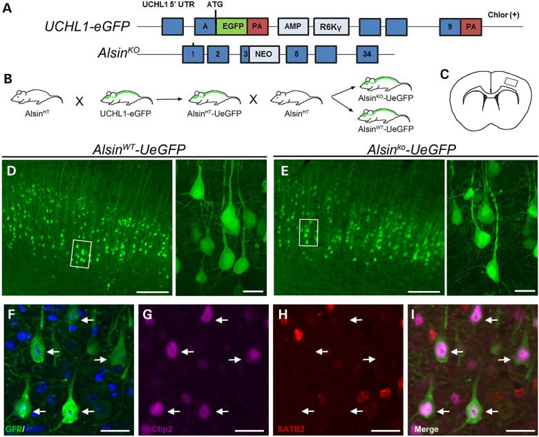
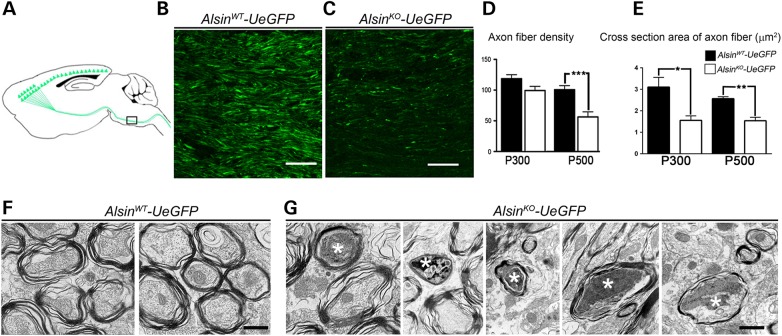
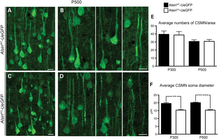
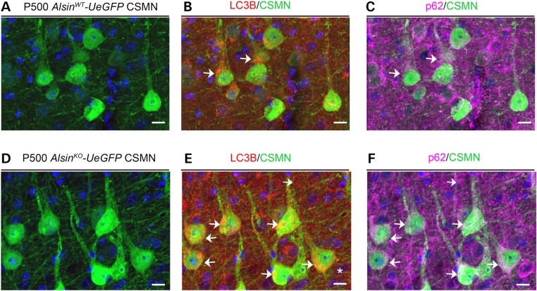
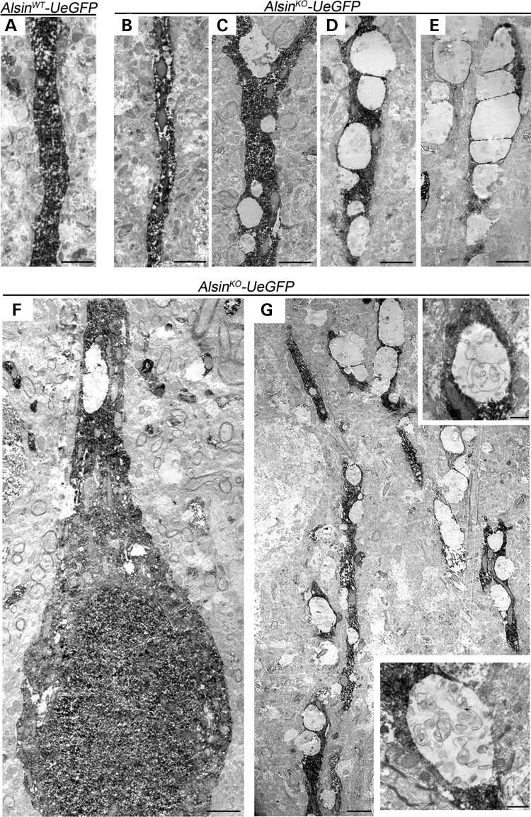
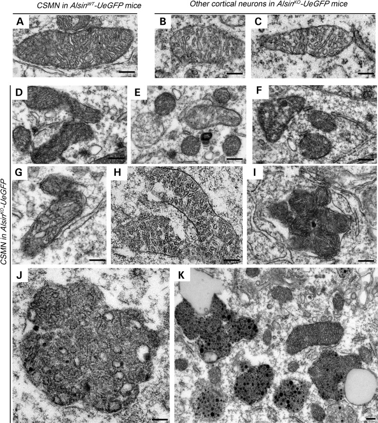
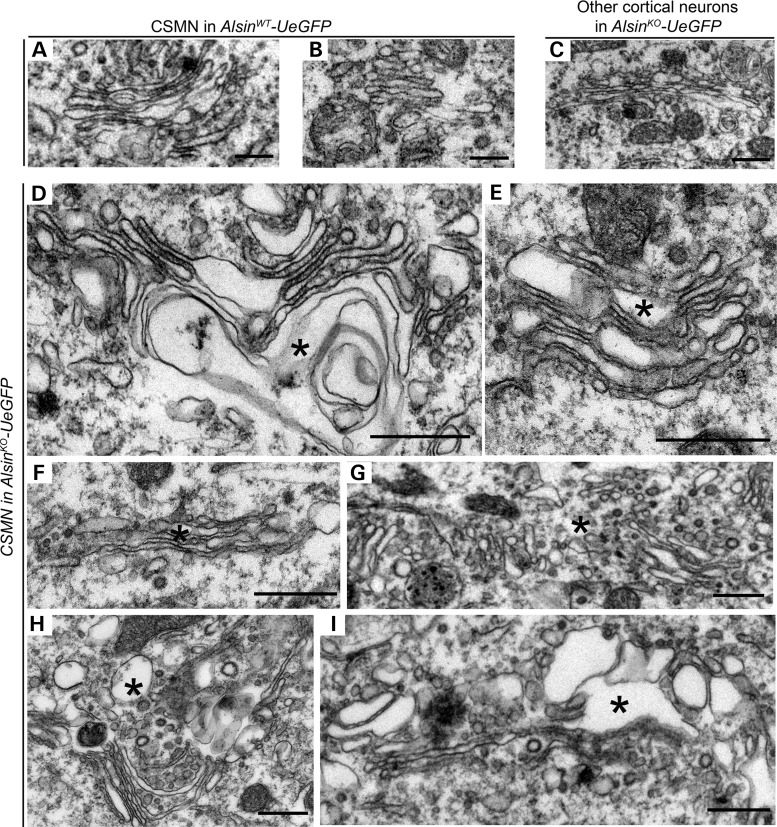
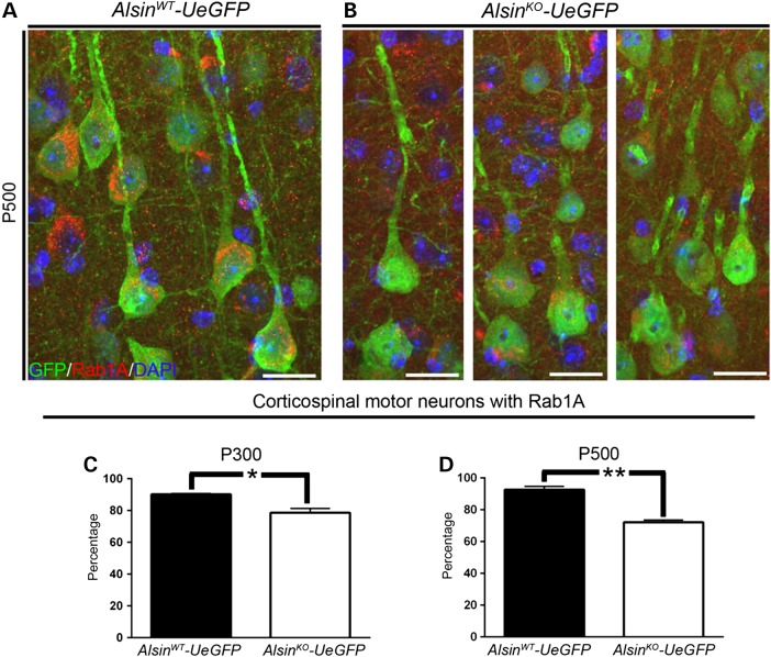
Mutations in the ALS2 gene result in early-onset amyotrophic lateral sclerosis, infantile-onset ascending hereditary spastic paraplegia and juvenile primary lateral sclerosis, suggesting prominent upper motor neuron involvement. However, the importance of alsin function for corticospinal motor neuron (CSMN) health and stability remains unknown. To date, four separate alsin knockout (Alsin(KO)) mouse models have been generated, and despite hopes of mimicking human pathology, none displayed profound motor function defects. This, however, does not rule out the possibility of neuronal defects within CSMN, which is not easy to detect in these mice. Detailed cellular analysis of CSMN has been hampered due to their limited numbers and the complex and heterogeneous structure of the cerebral cortex. In an effort to visualize CSMN in vivo and to investigate precise aspects of neuronal abnormalities in the absence of alsin function, we generated Alsin(KO)-UeGFP mice, by crossing Alsin(KO) and UCHL1-eGFP mice, a CSMN reporter line. We find that CSMN display vacuolated apical dendrites with increased autophagy, shrinkage of soma size and axonal pathology even in the pons region. Immunocytochemistry coupled with electron microscopy reveal that alsin is important for maintaining cellular cytoarchitecture and integrity of cellular organelles. In its absence, CSMN displays selective defects both in mitochondria and Golgi apparatus. UCHL1-eGFP mice help understand the underlying cellular factors that lead to CSMN vulnerability in diseases, and our findings reveal unique importance of alsin function for CSMN health and stability.


Figures

Figure 1.
Figure 1.
Neurons expressing eGFP in AlsinWT-UeGFP and AlsinKO-UeGFP mice retain CSMN identity. (A) Schematic representation of the transgenic constructs of UCHL1-eGFP and AlsinKO gene used to generate AlsinWT-UeGFP and AlsinKO-UeGFP mice, respectively. (B) Breeding strategy to generate AlsinWT-UeGFP and AlsinKO-UeGFP mice, reporter line of CSMN in the AlsinKO background. (C) A cross-section area of the brain. Boxed area represents layer V of motor cortex. (D and E) eGFP+ neurons reside in layer V of the motor cortex and display pyramidal neuron morphology with prominent apical dendrites in both AlsinWT-UeGFP (D) and AlsinKO-UeGFP (E), inset are enlarged to the right. (FI) eGFP+ neurons express Ctip2 (G) but not Satb2 (H). Scale bar: 500 µm (D and E), 20 µm (insets, F–I).
Figure 2.
Figure 2.
Corticospinal tract axons degenerate in AlsinKO-UeGFP mice. (A) Schematic drawing of sagittal section showing CST axons. Pons, (the boxed area), is used for quantification of axon fiber density and thickness. (B and C) Representative images of eGFP+ CST axons in pons of AlsinWT-UeGFP (B) and AlsinKO-UeGFP (C) mice. (D) The average fiber density is reduced in AlsinKO-UeGFP mice at P500, but not at P300. (E) The average cross-section area of axon fiber is gradually reduced in AlsinKO-UeGFP mice at P300 and P500. (F) Electron micrographs of CST axons in ventral pons of AlsinWT-UeGFP mice show healthy axon fiber bundles. (G) There are numerous degenerating axon fibers within CST of AlsinKO-UeGFP mice at P500 displaying a wide variety of morphological defects such as: membranous debris, mitochondria aggregation, collapsed synaptic vesicles and membrane debris, electrodense material, crenated mitochondria and final stages of degeneration (denoted by stars). Bar graph values are mean ± SEM. Student's t-test, ***P < 0.0001. Scale bar: 50 µm (B and C); 500 nm (F–G).
Figure 3.
Figure 3.
Reduced soma diameter suggests potential neuronal defects, without major cell loss. (A and B) Representative images of CSMN in AlsinWT-UeGFP and (C and D) AlsinKO-UeGFP mice at P500. (E) Average numbers of CSMN were comparable between AlsinWT-UeGFP and AlsinKO-UeGFP mice. (F) The average CSMN soma diameter significantly decreased in AlsinKO-UeGFP mice at P300 and at P500. Bar graph values are mean ± SEM. Student's t-test, ** P < 0.003; *** P < 0.0007. Scale bar: 20 µm (A–D).
Figure 4.
Figure 4.
CSMN display increased autophagy in the absence of alsin function. (AF) Representative images of CSMN expressing LC3B, a prominent autophagy marker, in AlsinWT-UeGFP (A) and AlsinKO-UeGFP (B–F) mice. (G and H) Quantitative analysis of percent CSMN with autophagy within their apical dendrites (G) and soma (H). Bar graph values are mean ± SEM. Student's t-test, *P < 0.02. Scale bar: 20 µm (A and B).
Figure 5.
Figure 5.
p62 is present in CSMN that expresses LC3B in AlsinKO-UeGFP mice (AC) Representative images of CSMN (A) that express LC3B (B) and p62 (C) in the AlsinWT-UeGFP mice. (DF) Representative images of CSMN (D) that express LC3B (E) and p62 (F) in the AlsinKO-UeGFP mice. Arrows indicate CSMN that express both LC3B and p62. Scale Bar: 10 µm.
Figure 6.
Figure 6.
eGFP immunocytochemistry coupled with EM reveal major defects within apical dendrites of diseased CSMN. (A) Apical dendrite of CSMN in AlsinWT-UeGFP mice are devoid of any vacuoles and display healthy morphology. (BE) Apical dendrites of CSMN in AlsinKO-UeGFP mice have vacuoles of different sizes and various stages. (F) A GFP labeled CSMN soma with vacuolated proximal apical dendrite and (G) distal apical dendrite with many vacuoles only in diseased CSMN. Insets display magnified view of representative vacuoles with numerous disintegrating cellular debris. Scale bar: 2 µm (A–G); 500 nm (insets).
Figure 7.
Figure 7.
Absence of alsin function leads to mitochondrial defects in CSMN. (AC) Representative images of mitochondria present in the CSMN of AlsinWT-UeGFP mice (A) and other cortical neurons of AlsinKO-UeGFP mice (B and C). (DK) Representative images of mitochondria present in the CSMN of AlsinKO-UeGFP mice. They display numerous structural defects, such as disintegration of inner membranes (D–G), broken cristae (H), fusion with other mitochondria (I), aggregation (J) and fusion of these aggregates with endolysosome-like structures (K). Scale bar: 200 nm (A–J); 1 µm (K).
Figure 8.
Figure 8.
Alsin function is required for the stability of Golgi apparatus in CSMN. (A–C) Representative images of Golgi apparatus present in the CSMN of AlsinWT-UeGFP mice (A) and other cortical neurons of AlsinKO-UeGFP mice (B and C). (DI) Representative images of Golgi apparatus present in the CSMN of AlsinKO-UeGFP mice. They display numerous structural defects, such as vacuolated cisternae (denoted by asterisk) (D and E), cisternae filled with dark material (F and G) and dark vesicles and vacuoles disrupting their ultrastructure (H and I). Scale bar: 500 nm (A–I).
Figure 9.
Figure 9.
Rab1A expression is reduced in diseased CSMN. (A and B) Representative images of Rab1A expression in CSMN of AlsinWT-UeGFP (A) and AlsinKO-UeGFP mice (B). (C and D) Percentage of CSMN with Rab1A is progressively reduced in the absence of alsin function at P300 (C) and P500 (D). Bar graph values are mean ± SEM. Student's t-test, *P < 0.01, **P < 0.001. Scale bar: 20 µm (A and B).

References

    1. Yang Y., Hentati A., Deng H.X., Dabbagh O., Sasaki T., Hirano M., Hung W.Y., Ouahchi K., Yan J., Azim A.C. et al. (2001) The gene encoding alsin, a protein with three guanine-nucleotide exchange factor domains, is mutated in a form of recessive amyotrophic lateral sclerosis. Nat. Genet., 29, 160–165. - PubMed
    1. Eymard-Pierre E., Lesca G., Dollet S., Santorelli F.M., di Capua M., Bertini E., Boespflug-Tanguy O. (2002) Infantile-onset ascending hereditary spastic paralysis is associated with mutations in the alsin gene. Am. J. Hum. Genet., 71, 518–527. - PMC - PubMed
    1. Hadano S., Hand C.K., Osuga H., Yanagisawa Y., Otomo A., Devon R.S., Miyamoto N., Showguchi-Miyata J., Okada Y., Singaraja R. et al. (2001) A gene encoding a putative GTPase regulator is mutated in familial amyotrophic lateral sclerosis 2. Nat. Genet., 29, 166–173. - PubMed
    1. Eker H.K., Unlu S.E., Al-Salmi F., Crosby A.H. (2014) A novel homozygous mutation in ALS2 gene in four siblings with infantile-onset ascending hereditary spastic paralysis. Eur. J. Med. Genet., 57, 275–278. - PubMed
    1. Wakil S.M., Ramzan K., Abuthuraya R., Hagos S., Al-Dossari H., Al-Omar R., Murad H., Chedrawi A., Al-Hassnan Z.N., Finsterer J. et al. (2014) Infantile-onset ascending hereditary spastic paraplegia with bulbar involvement due to the novel ALS2 mutation c.2761C>T. Gene, 536, 217–220. - PubMed

Publication types

MeSH terms

Substances

Supplementary concepts