

Left ventricular function assessment in cirrhosis: Current methods and future directions
- PMID: 26755864
- PMCID: PMC4698479
- DOI: 10.3748/wjg.v22.i1.112
Left ventricular function assessment in cirrhosis: Current methods and future directions
Abstract
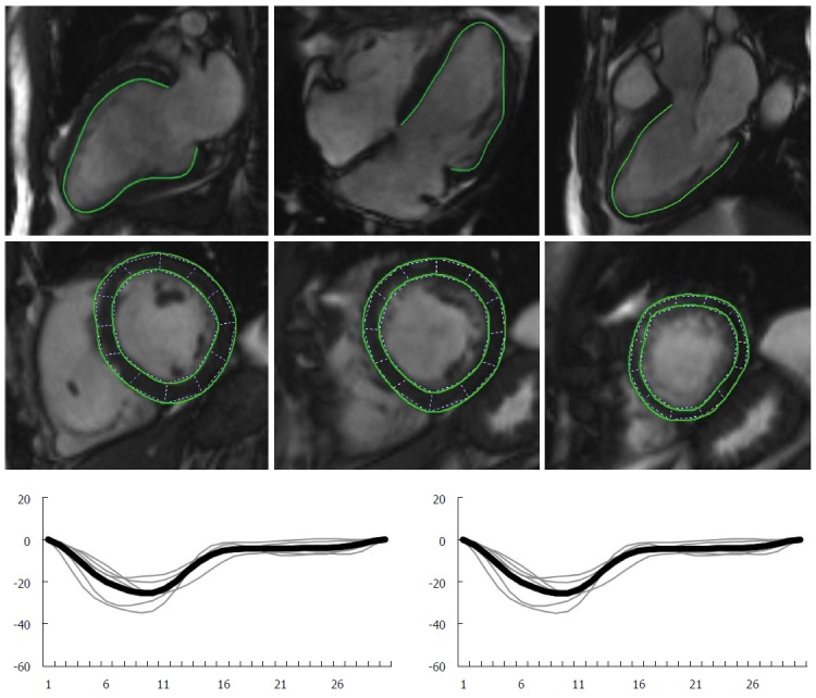
Cirrhotic cardiomyopathy has been defined as a chronic cardiac dysfunction in patients with cirrhosis characterized by impaired contractile responsiveness to stress and/or altered diastolic relaxation with electrophysiological abnormalities in the absence of other known cardiac disease. Non-invasive cardiovascular imaging modalities play a major role in unmasking systolic and diastolic dysfunction in patients with cirrhosis. Echocardiography has been the most commonly used modality for assessing myocardial function in these patients. Conventional echocardiographic indices rely on several assumptions that may limit their applicability in patients with a hyperdynamic circulation. Newer imaging modalities may contribute to a more accurate diagnosis of cardiovascular abnormalities in cirrhotic patients, thereby influencing clinical management. We aimed to review the different non-invasive imaging technologies currently used for assessing left ventricular systolic and diastolic function in cirrhosis, as well as to describe new imaging modalities with potential clinical applicability in the near future.
Keywords: Cardiomyopathy; Cirrhosis; Deformation imaging; Diastolic function; Echocardiography; Magnetic resonance imaging; Systolic function.
Figures


Similar articles
-
Systolic and diastolic dysfunction in cirrhosis: a tissue-Doppler and speckle tracking echocardiography study.Liver Int. 2013 Sep;33(8):1158-65. doi: 10.1111/liv.12187. Epub 2013 Apr 25. Liver Int. 2013. PMID: 23617332
-
Pathophysiological and clinical approach to cirrhotic cardiomyopathy.J Gastrointestin Liver Dis. 2014 Sep;23(3):301-10. doi: 10.15403/jgld.2014.1121.233.apac. J Gastrointestin Liver Dis. 2014. PMID: 25267959 Review.
-
LEFT ventricular function assessed by echocardiography in cirrhosis: relationship to systemic hemodynamics and renal dysfunction.J Hepatol. 2013 Jan;58(1):51-7. doi: 10.1016/j.jhep.2012.08.027. Epub 2012 Sep 16. J Hepatol. 2013. PMID: 22989573
-
An update on cirrhotic cardiomyopathy.Expert Rev Gastroenterol Hepatol. 2019 May;13(5):497-505. doi: 10.1080/17474124.2019.1587293. Epub 2019 Mar 8. Expert Rev Gastroenterol Hepatol. 2019. PMID: 30802157 Review.
-
Cardiac imaging in patients with chronic liver disease.Clin Physiol Funct Imaging. 2017 Jul;37(4):347-356. doi: 10.1111/cpf.12311. Epub 2015 Nov 5. Clin Physiol Funct Imaging. 2017. PMID: 26541640 Review.
Cited by
-
NT-proBNP and Echocardiographic Parameters in Liver Cirrhosis - Correlations with Disease Severity.Med Princ Pract. 2019 Apr 16;28(5):432-41. doi: 10.1159/000499930. Online ahead of print. Med Princ Pract. 2019. PMID: 30995644 Free PMC article.
-
Is Cirrhotic Cardiomyopathy Related to Cirrhosis Severity?Rambam Maimonides Med J. 2023 Jan 29;14(1):e0001. doi: 10.5041/RMMJ.10488. Rambam Maimonides Med J. 2023. PMID: 36719669 Free PMC article.
-
The Value of Myocardial Energy Expenditure in the Diagnosis of Cirrhotic Cardiomyopathy.Int J Gen Med. 2025 Jul 5;18:3767-3774. doi: 10.2147/IJGM.S529946. eCollection 2025. Int J Gen Med. 2025. PMID: 40636287 Free PMC article.
References
-
- Shorr E, Zweifach BW, Furchgott RF, Baez S. Hepatorenal factors in circulatory homeostasis. IV. Tissue origins of the vasotropic principles, VEM and VDM, which appear during evolution of hemorrhagi and tourniquet shock. Circulation. 1951;3:42–79. - PubMed
-
- Zardi EM, Abbate A, Zardi DM, Dobrina A, Margiotta D, Van Tassell BW, Afeltra A, Sanyal AJ. Cirrhotic cardiomyopathy. J Am Coll Cardiol. 2010;56:539–549. - PubMed
-
- Alqahtani SA, Fouad TR, Lee SS. Cirrhotic cardiomyopathy. Semin Liver Dis. 2008;28:59–69. - PubMed
-
- Møller S, Henriksen JH. Cardiovascular complications of cirrhosis. Gut. 2008;57:268–278. - PubMed
Publication types
MeSH terms
LinkOut - more resources
Full Text Sources
Other Literature Sources
Medical
