

The Effect of Alendronate on Various Graft Materials Used in Maxillary Sinus Augmentation: A Rabbit Study
- PMID: 26756022
- PMCID: PMC4706848
- DOI: 10.5812/ircmj.33569
The Effect of Alendronate on Various Graft Materials Used in Maxillary Sinus Augmentation: A Rabbit Study
Abstract
Background: Increasing sinus pneumatization and the accompanying alveolar bone resorption complicate dental implant placement. This problem can be overcome today by raising the maxillary sinus floor with graft materials. Bisphosphonates are commonly used to accelerate the recovery of the graft materials and to prevent resorption.
Objectives: The purpose of this study is to investigate whether systemic administration of a bisphosphonate (alendronate) would improve new bone formation and reduce fibrous tissue formation over a 6-week follow-up in rabbits treated with two different grafting materials for maxillary sinus floor augmentation.
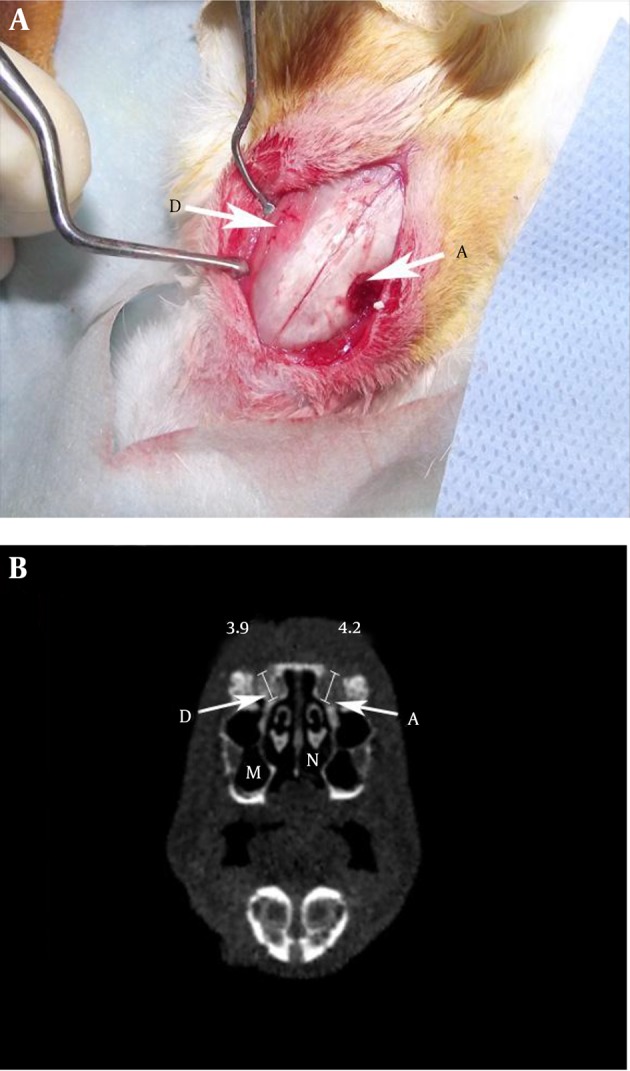
Materials and methods: This experimental animal study was conducted at the Experimental Medical Application and Research Center at Erzurum/ Turkey. Twelve New Zealand rabbits, each weighing between 2.7 and 3.3 kg, were used. Twenty-four maxillary sinus floor elevation operations were performed, two on each animal (n = 24). Each elevation was repaired with either deproteinized bovine bone (xenograft) or autogenous bone graft obtained from the iliac crest. Both groups were divided into 2 subgroups: saline-treated and alendronate-treated. All groups underwent the same surgical procedures and evaluation, and were sacrificed at the 6th postoperative week. Sinuses augmented with deproteinized bovine bone (xenograft) and autogenous bone graft were examined histopathologically and histomorphometrically.
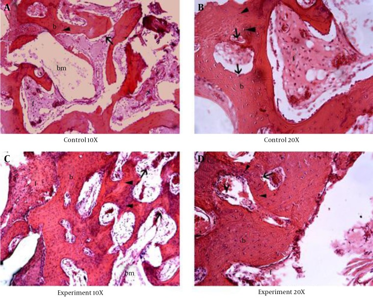
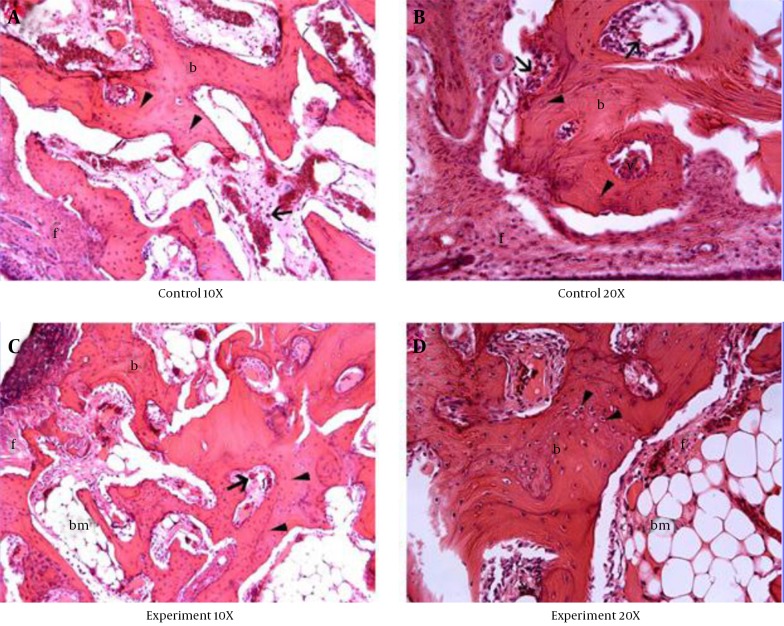
Results: At 6 weeks, the bone area was significantly larger in the Xenograft-Alendronate group (33.0% ± 5.0%) than in the Xenograft-Saline group (20.8% ± 4.9%) and the bone area was significantly larger in the Autogenous-Alendronate group (43.3% ± 3.8%) than in the Autogenous-Saline group (37.5% ± 6.6%) (P = 0.001). The histomorphometric and histopathological results consistently showed that alendronate stimulated bone formation and reduced fibrous tissue formation in maxillary sinus augmentation grafts, especially in the deproteinized bovine bone group (xenograft).
Conclusions: Alendronate may be considered a therapeutic option for improving the bone formation process and reducing resorption in different bone grafting procedures. Further detailed studies should focus on dosage and time-dependent effects of alendronate on bone remodeling.
Keywords: Alendronate; Bisphosphonate; Maxillary; Sinus.
Figures
Similar articles
-
Apical and marginal bone alterations around implants in maxillary sinus augmentation grafted with autogenous bone or bovine bone material and simultaneous or delayed dental implant positioning.Clin Oral Implants Res. 2011 May;22(5):485-91. doi: 10.1111/j.1600-0501.2010.02030.x. Epub 2010 Nov 19. Clin Oral Implants Res. 2011. PMID: 21087315
-
Effect of platelet-rich plasma with autogenous bone graft for maxillary sinus augmentation in a rabbit model.J Oral Maxillofac Surg. 2005 Mar;63(3):370-6. doi: 10.1016/j.joms.2004.07.017. J Oral Maxillofac Surg. 2005. PMID: 15742289
-
Rabbit maxillary sinus augmentation model with simultaneous implant placement: differential responses to the graft materials.J Periodontal Implant Sci. 2012 Dec;42(6):204-11. doi: 10.5051/jpis.2012.42.6.204. Epub 2012 Dec 31. J Periodontal Implant Sci. 2012. PMID: 23346463 Free PMC article.
-
Maxillary Sinus Floor Augmentation with Autogenous Bone Graft Alone Compared with Alternate Grafting Materials: a Systematic Review and Meta-Analysis Focusing on Histomorphometric Outcome.J Oral Maxillofac Res. 2020 Nov 30;11(3):e2. doi: 10.5037/jomr.2020.11302. eCollection 2020 Jul-Sep. J Oral Maxillofac Res. 2020. PMID: 33262881 Free PMC article. Review.
-
Maxillary Sinus Floor Augmentation with Autogenous Bone Graft Compared with a Composite Grafting Material or Bone Substitute Alone: a Systematic Review and Meta-Analysis Assessing Volumetric Stability of the Grafting Material.J Oral Maxillofac Res. 2021 Mar 31;12(1):e1. doi: 10.5037/jomr.2021.12101. eCollection 2021 Jan-Mar. J Oral Maxillofac Res. 2021. PMID: 33959236 Free PMC article. Review.
Cited by
-
Tomographic study of Jaw bone changes in patients with bisphosphonate-related osteonecrosis.J Clin Exp Dent. 2020 Mar 1;12(3):e285-e290. doi: 10.4317/jced.56265. eCollection 2020 Mar. J Clin Exp Dent. 2020. PMID: 32190200 Free PMC article.
-
Angulated Dental Implants in Posterior Maxilla FEA and Experimental Verification.Open Access Maced J Med Sci. 2018 Feb 14;6(2):397-401. doi: 10.3889/oamjms.2018.077. eCollection 2018 Feb 15. Open Access Maced J Med Sci. 2018. PMID: 29531612 Free PMC article.
-
Exploring the multifaceted impact of bisphosphonates on bone graft integration: transitioning from in vivo insights to clinical applications.Arch Toxicol. 2025 May;99(5):2157-2178. doi: 10.1007/s00204-025-03991-8. Epub 2025 Mar 6. Arch Toxicol. 2025. PMID: 40047864 Review.
References
-
- Wada K, Niimi A, Watanabe K, Sawai T, Ueda M. Maxillary sinus floor augmentation in rabbits: a comparative histologic-histomorphometric study between rhBMP-2 and autogenous bone. Int J Periodontics Restorative Dent. 2001;21(3):252–63. - PubMed
-
- Wanschitz F, Figl M, Wagner A, Rolf E. Measurement of volume changes after sinus floor augmentation with a phycogenic hydroxyapatite. Int J Oral Maxillofac Implants. 2006;21(3):433–8. - PubMed
-
- Tatum Jr H. Maxillary and sinus implant reconstructions. Dent Clin North Am. 1986;30(2):207–29. - PubMed
-
- Nkenke E, Radespiel-Troger M, Wiltfang J, Schultze-Mosgau S, Winkler G, Neukam FW. Morbidity of harvesting of retromolar bone grafts: a prospective study. Clin Oral Implants Res. 2002;13(5):514–21. - PubMed
LinkOut - more resources
Full Text Sources
Other Literature Sources