Review
. 2015 Dec 15;6(1):423-41.
doi: 10.1002/cphy.c150009.

Cytochrome P450 and Lipoxygenase Metabolites on Renal Function

Affiliations
Review

Cytochrome P450 and Lipoxygenase Metabolites on Renal Function

John D Imig et al. Compr Physiol. .

Abstract

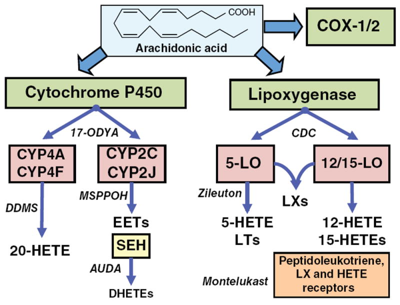
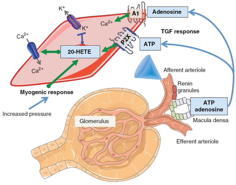
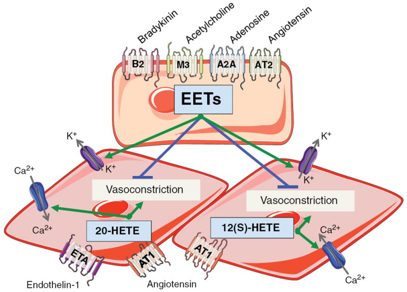
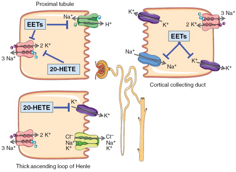
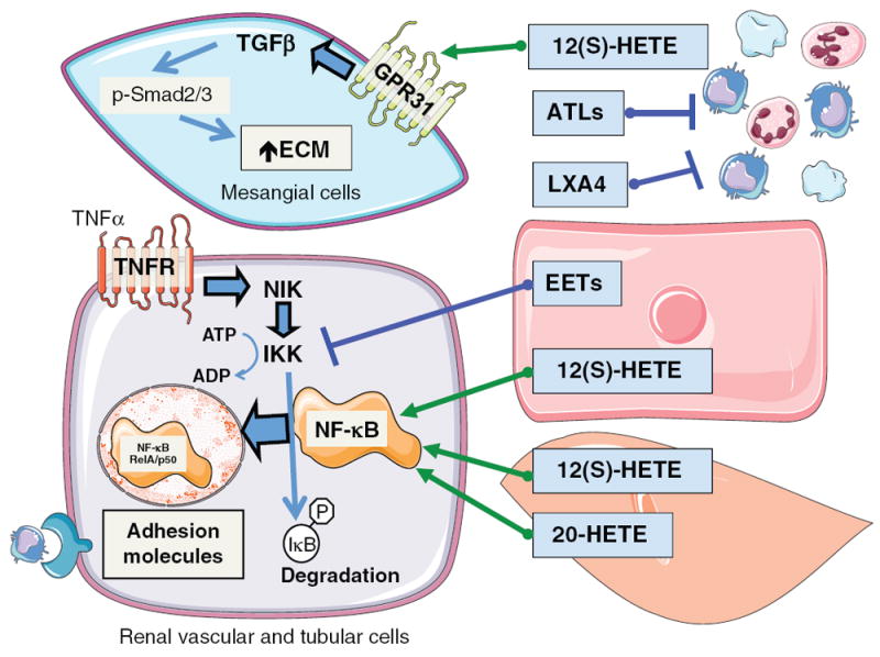
Arachidonic acid metabolites have a myriad of biological actions including effects on the kidney to alter renal hemodynamics and tubular transport processes. Cyclooxygenase metabolites are products of an arachidonic acid enzymatic pathway that has been extensively studied in regards to renal function. Two lesser-known enzymatic pathways of arachidonic acid metabolism are the lipoxygenase (LO) and cytochrome P450 (CYP) pathways. The importance of LO and CYP metabolites to renal hemodynamics and tubular transport processes is now being recognized. LO and CYP metabolites have actions to alter renal blood flow and glomerular filtration rate. Proximal and distal tubular sodium transport and fluid and electrolyte homeostasis are also significantly influenced by renal CYP and LO levels. Metabolites of the LO and CYP pathways also have renal actions that influence renal inflammation, proliferation, and apoptotic processes at vascular and epithelial cells. These renal LO and CYP pathway actions occur through generation of specific metabolites and cell-signaling mechanisms. Even though the renal physiological importance and actions for LO and CYP metabolites are readily apparent, major gaps remain in our understanding of these lipid mediators to renal function. Future studies will be needed to fill these major gaps regarding LO and CYP metabolites on renal function.


Figures

Figure 1
Figure 1
Diagram depicting pathways of arachidonic metabolism. CYP, LO, and COX, pathways can metabolize arachidonic acid. The contribution of COX-1 and COX-2 metabolites to renal function has been extensively reviewed (13, 78). CYP enzymes can generate 20-HETE and EETs. sEH can hydrate EETs to form dihydroxyeicosatrienoic acids (DHETEs). LO enzymes generate HETEs, leukotrienes (LTs) and lipoxins (LXs). Representative enzymatic inhibitors and receptor antagonists are named in italics.
Figure 2
Figure 2
Diagram depicting renal blood flow autoregulatory tubuloglomerular feedback (TGF) and myogenic responses. Afferent arteriolar vascular smooth muscle cell depicts contribution of 20-HETE to myogenic and TGF responses. Increased afferent arteriolar transmural pressure triggers the myogenic response to increase 20-HETE levels. 20-HETE causes afferent arteriolar constriction via inhibition of large-conductance Ca2+-activated K+ channels and activation of L-type Ca2+ channels. TGF response is initiated by sensing cells at the macula densa resulting in release of adenosine and ATP. Adenosine activates adenosine type A1 receptors to increase intracellular Ca2+ and ATP activates purinergic P2X receptors to increase 20-HETE levels resulting in afferent arteriolar constriction.
Figure 3
Figure 3
Diagram depicting renal vascular endothelial cell and vascular smooth muscle cell signaling for EETs, 20-HETE, and 12(S)-HETE. Bradykinin B2 receptor, acetylcholine muscarinic M3 receptor, adenosine A2A receptor, or AT2 receptor activation increases renal endothelial cell EET levels. EETs released from the endothelial cell act in a paracrine manner on vascular smooth muscle cells to activate large-conductance Ca2+-activated K+ channels and oppose vasoconstriction. ETA receptor activation increases renal vascular smooth muscle cell 20-HETE levels. AT1 receptor activation increases vascular smooth muscle cell 20-HETE and 12(S)-HETE activates. 20-HETE and 12(S)-HETE activate L-type Ca2+ channels and cause vasoconstriction.
Figure 4
Figure 4
Diagram depicting renal proximal tubule, TALH, and CCD cell signaling for EETs and 20-HETE. Proximal tubule cell: 20-HETE inhibits basolateral Na-K ATPase activity to decrease sodium reabsorption. EETs inhibit basolateral Na-K ATPase activity and apical NHE3 to decrease sodium reabsorption. TALH: 20-HETE inhibits apical K+ channels to limit K+ availability for transport via the Na-K-2Cl transporter resulting in decreased lumen positive transepithelial potential and passive reabsorption of cations. CCD: EETs inhibit apical ENaC and basolateral K+ channels to decrease sodium reabsorption.
Figure 5
Figure 5
Diagram depicting mesangial cell hypertrophic and renal vascular and tubular cell inflammatory responses to CYP and LO metabolites. Circulating platelets, and leukocytes can generate LO metabolites, 12(S)-HETE, ATLs, and LXA. 12(S)-HETE acts on glomerular mesangial cells to activate GPR31 receptors resulting in increased TGF-β resulting in phosphorylation of Smad2/3 to increase extracellular matrix and hypertrophy. ATLs and LXA4 interfere with activation and migration of inflammatory cells. Renal endothelial cells generate EETs, endothelial and vascular smooth muscle cells generate 12(S)-HETE and vascular smooth muscle cells generate 20-HETE to act in a paracrine manner to influence the renal vascular and tubular cell inflammatory responses. TNF-α acting on the TNF receptor activates a sequence of cell signaling events resulting in NF-κB translocation to the nucleus to increase adhesion molecule levels to cause inflammatory cell infiltration. EETs act to prevent IKK phosphorylation and activation of NF-κB to decrease renal inflammation. 12(S)-HETE and 20-HETE increase NF-κB activation and translocation to the nucleus to increase renal inflammation.

References

    1. Akbulut T, Regner KR, Roman RJ, Avner ED, Falck JR, Park F. 20-HETE activates the Raf/MEK/ERK pathway in renal epithelial cells through an EGFR- and c-Src-dependent mechanism. Am J Physiol Renal Physiol. 2009;297:F662–F670. - PMC - PubMed
    1. Athirakul K, Bradbury JA, Graves JP, DeGraff LM, Ma J, Zhao Y, Couse JF, Quigley R, Harder DR, Zhao X, Imig JD, Pedersen TL, Newman JW, Hammock BD, Conley AJ, Korach KS, Coffman TM, Zeldin DC. Increased blood pressure in mice lacking cytochrome P450 2J5. FASEB J. 2008;22:4096–4108. - PMC - PubMed
    1. Alonso-Galicia M, Drummond HA, Reddy KK, Falck JR, Roman RJ. Inhibition of 20-HETE production contributes to the vascular responses to nitric oxide. Hypertension. 1997;29:320–325. - PubMed
    1. Alonso-Galicia M, Falck JR, Reddy KM, Roman RJ. 20-HETE agonists and antagonists in the renal circulation. Am J Physiol. 1999;277:F790–F796. - PubMed
    1. Alonso-Galicia M, Maier KG, Greene AS, Cowley AW, Roman RJ. Role of 20-Hydroxyeicosatetraeinoic acid in the renal and vasoconstrictor actions of angiotensin II. Am J Physiol Regulatory, Integrative Comp Physiol. 2002;283:R60–R68. - PubMed

Publication types

Substances

LinkOut - more resources