Review
. 2015 Dec 15;6(1):507-26.
doi: 10.1002/cphy.c150022.

Mitochondrial Dynamics and Heart Failure

Affiliations
Review

Mitochondrial Dynamics and Heart Failure

A A Knowlton et al. Compr Physiol. .

Abstract

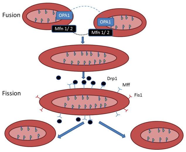
Mitochondrial dynamics, fission and fusion, were first identified in yeast with investigation in heart cells beginning only in the last 5 to 7 years. In the ensuing time, it has become evident that these processes are not only required for healthy mitochondria, but also, that derangement of these processes contributes to disease. The fission and fusion proteins have a number of functions beyond the mitochondrial dynamics. Many of these functions are related to their membrane activities, such as apoptosis. However, other functions involve other areas of the mitochondria, such as OPA1's role in maintaining cristae structure and preventing cytochrome c leak, and its essential (at least a 10 kDa fragment of OPA1) role in mtDNA replication. In heart disease, changes in expression of these important proteins can have detrimental effects on mitochondrial and cellular function.


Figures

Figure 1
Figure 1
Mitochondrial Fusion and Fission: Diagram summarizes mitochondrial fusion and fission. Fusion (top panels) - Mitofusin (Mfn) 1 and 2 together fuse the outer mitochondrial membrane. Expression of these two proteins is unchanged in HF. OPA1 fuses the inner mitochondrial membrane, leading to a single, larger mitochondrion, and OPA1 is decreased in HF. Fission (lower panels) - Fission divides one mitochondrion into two smaller mitochondria. Mff recruits Drp1 to the mitochondria for fission. Drp1 is a cytoplasmic protein, but forms complexes at fission sites on the outer mitochondrial mediating fission.Fis1’s tetratricopeptide repeat motif helps create a scaffold, which facilitates the formation of protein clusters on the outer mitochondrial membrane, but is not essential for fission, as Drp1 can complex on the mitochondrial surface without Fis1, if Mff is present. Mitochondrial fission and fusion are essential for maintenance of normal mitochondria. A key aspect of fusion in the heart may be asymetric fission, as described by Twig et al. (see text). Asymmetric fission generates a smaller mitochondrion with a decreased Δψ m and a larger mitochondrion with a preserved Δψ m. The small, depolarized mitochondrion is removed by mitophagy. Figure updated from Knowlton and Chen (88).
Figure 2
Figure 2
Mitochondrial Protein Trafficking: Diagram summarizes key pathways for protein and solute import into the mitochondria. 1) The TIM/TOM complex is the major mitochondrial proteins importing machinery. Proteins that are encoded in the nucleus and synthesized in the cytoplasm are transported into the mitochondrion through TIM/TOM assisted by chaperones. 2) VDAC and ANT have roles in small solute trafficking, but in the stressed cell they are part of the mPTP, which opens leading mitochondrial depolarization. 3) mPTP, spanning the IMM and OMM, consists of membranous elements, including VDAC on the OMM, ANT on the IMM. Under conditions like high Ca+2 concentration, increased oxidative stress, low ATP, and mitochondrial depolarization, mPTP is allows free diffusion of solutes across the membranes, which ultimately leads to mitochondrial swelling, and apoptosis.
Figure 3
Figure 3
Mitophagy: Summary of steps involved in mitophagy. The depolarized mitochondrion leads to accumulation of PINK1, which phosphorylates Mfn2, which acts as a lure for Parkin. Parkin binding Mfn2 triggers mitophagy. Outer membrane proteins, including both Mfn’s and VDAC, are ubiquitinated. P62 is recruited, binding the ubiquitinated proteins, linking them to LC3. The isolation membrane elongtes and eventually engulfs the mitochondrial pieces destined for autophagy, forming the autophagosome, which eventually fuses with the lysosome, leading to degradation of the enclosed mitochondria.
Figure 4
Figure 4
Summary of Mitochondrial Dynamics and Ischemic Heart Failure. Major findings with multiple studies supporting them are shown. The next few years should lead to identification of more roles for fission and fusion in heart failure.

References

    1. Ahuja P, Wanagat J, Wang Z, Wang Y, Liem DA, Ping P, et al. Divergent Mitochondrial Biogenesis Responses in Human Cardiomyopathy. Circulation. 2013 Apr 15;127:1957–67. - PMC - PubMed
    1. Aich A, Shaha C. Novel Role of Calmodulin in Regulating Protein Transport to Mitochondria in a Unicellular Eukaryote. Molecular and Cellular Biology. 2013 Nov 15;33(22):4579–93. - PMC - PubMed
    1. Altmann R. Die Elementarorganismen und ihre Beziehungen zu den Zellen. Leipzig: Veit & Co; 1890.
    1. Amati-Bonneau P, Milea D, Bonneau D, Chevrollier A, Ferré M, Guillet V, et al. OPA1-associated disorders: Phenotypes and pathophysiology. The International Journal of Biochemistry & Cell Biology. 2009 Oct;41(10):1855–65. - PubMed
    1. Ambrosio G, Zweier JL, Duilio C, Kuppusamy P, Santoro G, Elia PP, et al. Evidence that mitochondrial respiration is a source of potentially toxic oxygen free radicals in intact rabbit hearts subjected to ischemia and reflow. Journal of Biological Chemistry. 1993 Sep 5;268(25):18532–41. - PubMed

Publication types