. 2016 Jan 12;11(1):e0146981.
doi: 10.1371/journal.pone.0146981. eCollection 2016.

Altered Circadian Food Anticipatory Activity Rhythms in PACAP Receptor 1 (PAC1) Deficient Mice

Affiliations

Altered Circadian Food Anticipatory Activity Rhythms in PACAP Receptor 1 (PAC1) Deficient Mice

Jens Hannibal et al. PLoS One. .

Abstract

Light signals from intrinsically photosensitive retinal ganglion cells (ipRGCs) entrain the circadian clock and regulate negative masking. Two neurotransmitters, glutamate and Pituitary Adenylate Cyclase Activating Polypeptide (PACAP), found in the ipRGCs transmit light signals to the brain via glutamate receptors and the specific PACAP type 1 (PAC1) receptor. Light entrainment occurs during the twilight zones and has little effect on clock phase during daytime. When nocturnal animals have access to food only for a few hours during the resting phase at daytime, they adapt behavior to the restricted feeding (RF) paradigm and show food anticipatory activity (FAA). A recent study in mice and rats demonstrating that light regulates FAA prompted us to investigate the role of PACAP/PAC1 signaling in the light mediated regulation of FAA. PAC1 receptor knock out (PAC1-/-) and wild type (PAC1+/+) mice placed in running wheels were examined in a full photoperiod (FPP) of 12:12 h light/dark (LD) and a skeleton photoperiod (SPP) 1:11:1:11 h L:DD:L:DD at 300 and 10 lux light intensity. Both PAC1-/- mice and PAC1+/+ littermates entrained to FPP and SPP at both light intensities. However, when placed in RF with access to food for 4-5 h during the subjective day, a significant change in behavior was observed in PAC1-/- mice compared to PAC1+/+ mice. While PAC1-/- mice showed similar FAA as PAC1+/+ animals in FPP at 300 lux, PAC1-/- mice demonstrated an advanced onset of FAA with a nearly 3-fold increase in amplitude compared to PAC1+/+ mice when placed in SPP at 300 lux. The same pattern of FAA was observed at 10 lux during both FPP and SPP. The present study indicates a role of PACAP/PAC1 signaling during light regulated FAA. Most likely, PACAP found in ipRGCs mediating non-image forming light information to the brain is involved.


Conflict of interest statement

Competing Interests: The authors have declared that no competing interests exist.

Figures

Fig 1
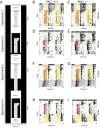
Fig 1. Food anticipatory activity (FAA) during restricted feeding (RF) and different light conditions.
A. Restricted feeding protocols with food deprivation during the daytime starting at ZT 4 in PAC1 +/+ and PAC1-/- mice were fed ad libitum and then the availability of food were gradually reduced (RF) from 6 h/day to 4 h/day (ZT4—ZT8). The RF time during each experiment (1–4) is indicated by gray shading, light in white and dark in black. Experiment 1 was performed with light in a full photoperiod (FPP) of 12:12 h light/darkness (L/D) at 300 lux. Experiment 2 was performed in a skeleton photoperiod (SPP) of 1:10:1:12 h L/D/L/D at 300 lux. Experiment 3 was performed with light in a full photoperiod (FPP) of 12:12 h L/D at 10 lux followed by a period of DD for 10 days. Experiment 4 was performed in a skeleton photoperiod (SPP) of 1:10:1:12 h L/D/L/D at 10 lux. Representative actograms from PAC1+/+ and PAC1-/- mice in Experiment 1 are shown in B-C, Experiment 2 in D-E, Experiment 3 in F-G, and Experiment 4 in H-I. In B-I RF is indicated by red shading, light periods by yellow shading and constant darkness in Experiment 3 by gray shading. The light-dark cycle is indicated by the white and black bars on top of each actogram, respectively.
Fig 2
Fig 2. Food anticipatory activity (FAA) in PAC1+/+ and PAC1-/- receptor deficient mice under a full photoperiod (FPP) and skeleton photoperiod (SPP) at light intensities of either 300 lux or 10 lux.
The light/dark period is indicated below the X-axis in yellow/black. The same 8 animals of each genotype were used in all experiments and data are average of 5 d of activity (see also material and methods). A. Group mean (± SEM) waveform of wheel running activity of PAC1+/+ (blue line) and PAC1-/- mice (red line) during FPP at 300 lux. B. Group mean (± SEM) waveform of wheel running activity of the same animals as in A showing FAA before mealtime (indicated by light green). C. Group mean (± SEM) waveform of wheel running activity of PAC1+/+ (blue line) and PAC1 -/- mice (red line) during SPP at 300 lux. D. Group mean (± SEM) waveform of wheel running activity of the same animals as in C showing FAA before mealtime (indicated by light green). E and F correspond to A and B but at light intensity of 10 lux. G and H correspond to C and D but at light intensity of 10 lux.
Fig 3
Fig 3. A-D. Weight profiles (mean ± SEM) of during RF regime of PAC1+/+ (PAC1+/+) (blue line) and PAC1 (PAC1-/-) receptor deficient mice (red line) shown in Fig 2.
Start weight was set to 100% and the mean of the relative weight change every second day during RF for each animal of the two groups was plotted.

References

    1. Mohawk JA, Takahashi JS. Cell autonomy and synchrony of suprachiasmatic nucleus circadian oscillators. Trends Neurosci. 2011;34(7):10. - PMC - PubMed
    1. Mohawk JA, Green CB, Takahashi JS. Central and peripheral circadian clocks in mammals. Annu Rev Neurosci. 2012;35:445–62. 10.1146/annurev-neuro-060909-153128 - DOI - PMC - PubMed
    1. Golombek DA, Rosenstein RE. Physiology of circadian entrainment. Physiol Rev. 2010;90(3):1063–102. 10.1152/physrev.00009.2009 - DOI - PubMed
    1. Sand A, Schmidt TM, Kofuji P. Diverse types of ganglion cell photoreceptors in the mammalian retina. Prog Retin Eye Res. 2012. - PMC - PubMed
    1. Do MT, Yau KW. Intrinsically photosensitive retinal ganglion cells. Physiol Rev. 2010;90(4):1547–81. 10.1152/physrev.00013.2010 - DOI - PMC - PubMed

Publication types

MeSH terms

Substances