. 2016 Mar 11;291(11):5676-5687.
doi: 10.1074/jbc.M115.711689. Epub 2016 Jan 12.

Novel Features of Eukaryotic Photosystem II Revealed by Its Crystal Structure Analysis from a Red Alga

Affiliations

Novel Features of Eukaryotic Photosystem II Revealed by Its Crystal Structure Analysis from a Red Alga

Hideo Ago et al. J Biol Chem. .

Abstract

Photosystem II (PSII) catalyzes light-induced water splitting, leading to the evolution of molecular oxygen indispensible for life on the earth. The crystal structure of PSII from cyanobacteria has been solved at an atomic level, but the structure of eukaryotic PSII has not been analyzed. Because eukaryotic PSII possesses additional subunits not found in cyanobacterial PSII, it is important to solve the structure of eukaryotic PSII to elucidate their detailed functions, as well as evolutionary relationships. Here we report the structure of PSII from a red alga Cyanidium caldarium at 2.76 Å resolution, which revealed the structure and interaction sites of PsbQ', a unique, fourth extrinsic protein required for stabilizing the oxygen-evolving complex in the lumenal surface of PSII. The PsbQ' subunit was found to be located underneath CP43 in the vicinity of PsbV, and its structure is characterized by a bundle of four up-down helices arranged in a similar way to those of cyanobacterial and higher plant PsbQ, although helices I and II of PsbQ' were kinked relative to its higher plant counterpart because of its interactions with CP43. Furthermore, two novel transmembrane helices were found in the red algal PSII that are not present in cyanobacterial PSII; one of these helices may correspond to PsbW found only in eukaryotic PSII. The present results represent the first crystal structure of PSII from eukaryotic oxygenic organisms, which were discussed in comparison with the structure of cyanobacterial PSII.

Keywords: crystal structure; crystallography; cyanobacteria; electron transfer complex; membrane protein; photosynthesis; photosystem II; x-ray crystallography.


Figures

FIGURE 1.
FIGURE 1.
Protein composition analyzed by SDS-PAGE of the red algal PSII. Lane 1, PSII before crystallization; lane 2, dissolved PSII crystals.
FIGURE 2.
FIGURE 2.
A part of the electron density map obtained from crystals of the red algal PSII tetramer at 2.76 Å resolution, showing the quality of the map and its fitting with the amino acid residues.
FIGURE 3.
FIGURE 3.
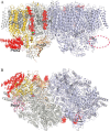
Crystal structure of red algal PSII and its comparison with cyanobacterial PSII. A, a tetramer of red algal PSII within a crystallographic asymmetric unit. The view is perpendicular to the membrane plane. The boxed areas represent two dimers, respectively, which are stacked in the stromal sides with a rotational angle of ∼20° with respect to each other. B, structure of a dimer (Mol-3/Mol-4) viewed from the lumenal side. The black dashed line indicate the intersurface between two monomers in a dimer. For clarity, the four extrinsic subunits and extrinsic loop regions of intrinsic subunits were omitted. The transmembrane helices of one monomer of the dimer are labeled. C, superimposition of the red algal PSII dimer with the cyanobacterial PSII dimer. Green represents the cyanobacterial dimer, and cyan and red represent Mol-1 and Mol-2 from a tetramer of the red algal PSII. The area circled with a black dashed line indicate the PsbQ′ subunit, which was not found in the cyanobacterial PSII.
FIGURE 4.
FIGURE 4.
A part of unit cell in the crystals of red algal PSII, showing the contacts among adjacent PSII dimers. A, view perpendicular to the membrane plane along with a PSII dimer in the middle. The circled areas represent PsbQ′, which is present in one side but absent in the opposite side of a dimer. B, same as A, but rotated by 180° along the membrane plane. PsbQ′ was colored in red, and PsbZ was colored in green.
FIGURE 5.
FIGURE 5.
Structure of red algal PSII dimer (Mol-1, right; Mol-2, left) and the location of PsbQ′ and two novel transmembrane helices. A, side view of the thylakoid membrane. The cycle with red dashed line represents the region where the PsbQ′ subunit was missing. B, top view from the stromal side of the membrane. The electron densities for PsbQ′ and the two novel helices (chains S and W) were depicted in red as omit maps contoured at 2 σ in both A and B.
FIGURE 6.
FIGURE 6.
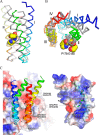
Structure of the red algal PsbQ′ protein, its comparison with the structure of spinach PsbQ, and its interactions with red algal CP43. A, superimposition of the structures of red algal PsbQ′ (colored) and spinach PsbQ (gray). The balls represent three residues (PGG) from CP43 (Pro191–Gly193) that are in contact with the helix II of PsbQ′ (for clarity, the two residues of CP43-Gly194 and CP43-Asp195 were omitted). B, top view of A. C, interactions of red algal PsbQ′ with CP43 depicted by the surface potential of the two subunits. Red represents negative, and blue represents positive potentials.
FIGURE 7.
FIGURE 7.
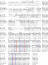
Alignment of the PsbQ′ homologous protein sequences from representative species of various groups of organisms available in the databases with the program Clustal Omega (41). The species selected include red algae (C. caldarium and Galdieria sulfuraria), a cyanobacterium (Synechocystis sp. PCC 6803), a cryptomonad (Guillardia theta), a diatom (Chaetoceros gracilis), a brown alga (Ectocarpus siliculosus), a green alga (Chlamydomonas reinhardtii), a moss (Physcomitrella patens), and two higher plants (Arabidopsis thaliana and Spinacia oleracea). Residues highlighted in red indicate completely conserved residues in all of the organisms, and areas boxed in blue indicate regions where the distance between Cα of the PsbQ′ protein and PSII intrinsic subunits are within 5 Å.
FIGURE 8.
FIGURE 8.
Alignment of a part of the CP43 protein sequences in the lumenal side where the region of interaction with the PsbQ′ protein is located, from representative species selected as in Fig. 7. The red boxed residues indicate the possible interaction sites with PsbQ′ revealed in the present study.
FIGURE 9.
FIGURE 9.
Structures and their environments of the two new transmembrane helices. A, chain S. B, chain W. Both are viewed from the side of the thylakoid membrane.

References

    1. Zouni A., Witt H. T., Kern J., Fromme P., Krauss N., Saenger W., and Orth P. (2001) Crystal structure of photosystem II from Synechococcus elongatus at 3.8 Angstrom resolution. Nature 409, 739–743 - PubMed
    1. Kamiya N., and Shen J.-R. (2003) Crystal structure of oxygen-evolving photosystem II from Thermosynechococcus vulcanus at 3.7-A resolution. Proc. Natl. Acad. Sci. U.S.A. 100, 98–103 - PMC - PubMed
    1. Ferreira K. N., Iverson T. M., Maghlaoui K., Barber J., and Iwata S. (2004) Architecture of the photosynthetic oxygen-evolving center. Science 303, 1831–1838 - PubMed
    1. Guskov A., Kern J., Gabdulkhakov A., Broser M., Zouni A., and Saenger W. (2009) Cyanobacterial photosystem II at 2.9 Å resolution and role of quinines, lipids, channels and chloride. Nat. Struct. Mol. Biol. 16, 334–342 - PubMed
    1. Umena Y., Kawakami K., Shen J.-R., and Kamiya N. (2011) Crystal structure of oxygen-evolving photosystem II at a resolution of 1.9 Å. Nature 473, 55–60 - PubMed

Publication types

MeSH terms

Substances

Associated data

LinkOut - more resources