. 2016 Jan 13:7:10347.
doi: 10.1038/ncomms10347.

Methylation-dependent regulation of HIF-1α stability restricts retinal and tumour angiogenesis

Affiliations

Methylation-dependent regulation of HIF-1α stability restricts retinal and tumour angiogenesis

Yunho Kim et al. Nat Commun. .

Abstract

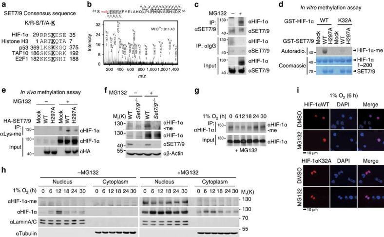
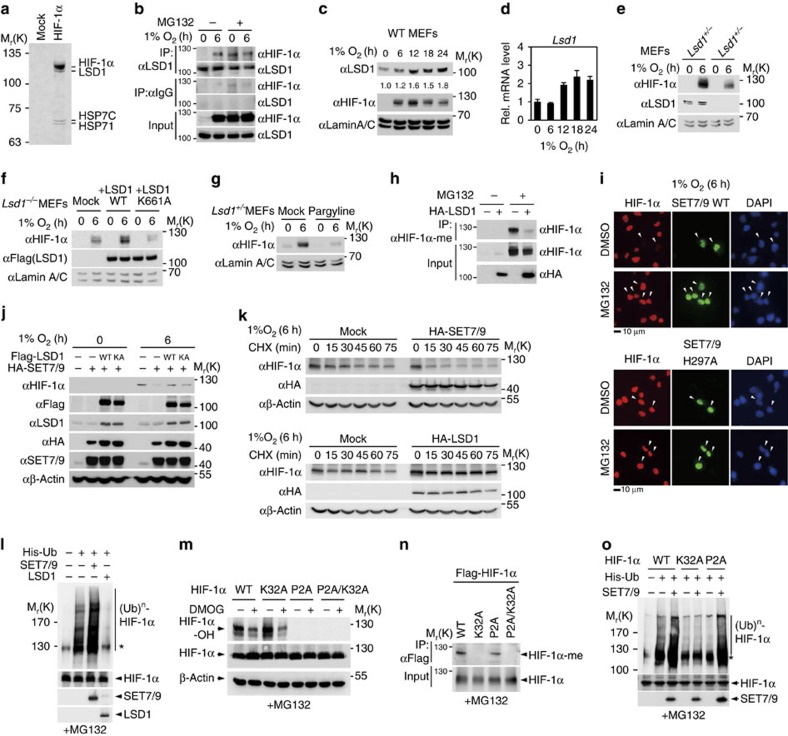
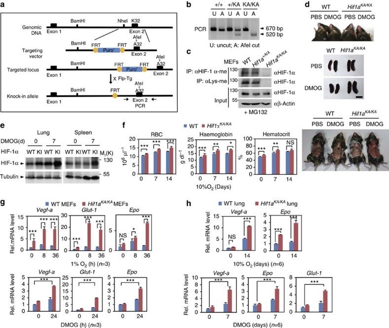
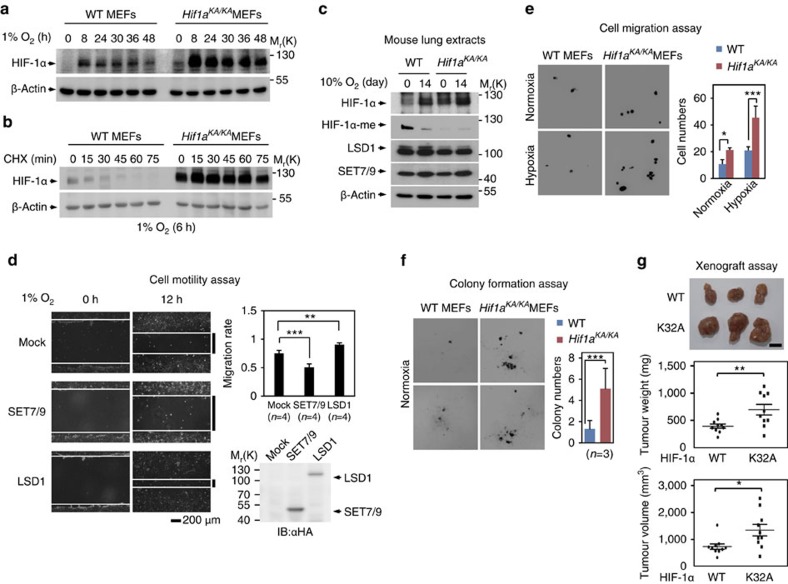
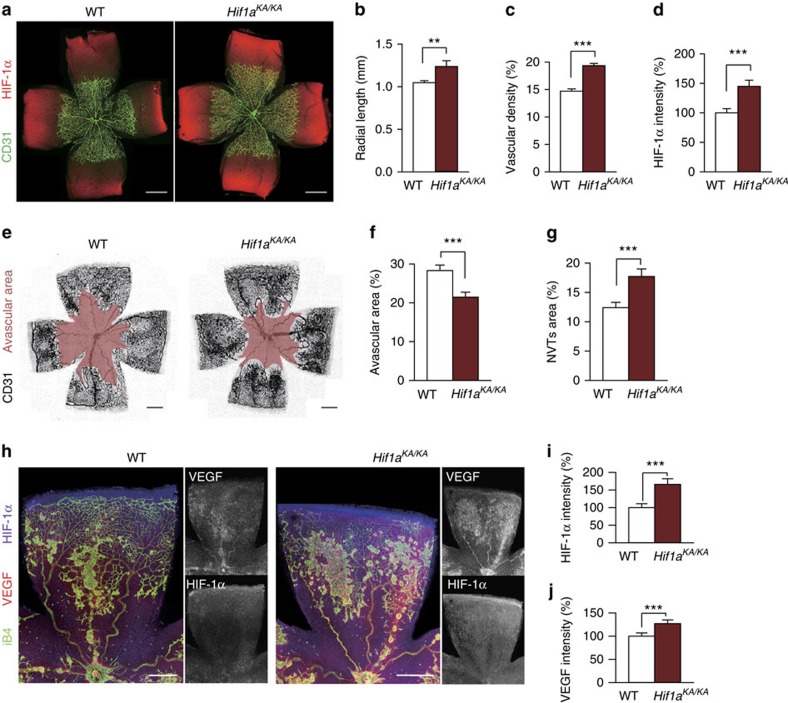
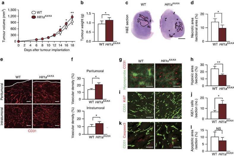
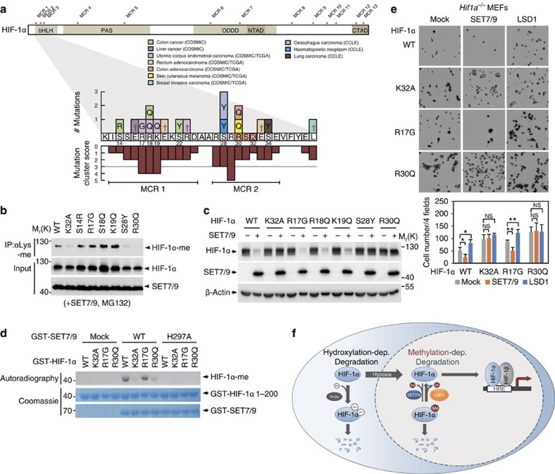
Hypoxia-inducible factor-1α (HIF-1α) mediates hypoxic responses and regulates gene expression involved in angiogenesis, invasion and metabolism. Among the various HIF-1α posttranslational modifications, HIF-1α methylation and its physiological role have not yet been elucidated. Here we show that HIF-1α is methylated by SET7/9 methyltransferase, and that lysine-specific demethylase 1 reverses its methylation. The functional consequence of HIF-1α methylation is the modulation of HIF-1α stability primarily in the nucleus, independent of its proline hydroxylation, during long-term hypoxic and normoxic conditions. Knock-in mice bearing a methylation-defective Hif1a(KA/KA) allele exhibit enhanced retinal angiogenesis and tumour vascularization via HIF-1α stabilization. Importantly, S28Y and R30Q mutations of HIF-1α, found in human cancers, are involved in the altered HIF-1α stability. Together, these results demonstrate a role for HIF-1α methylation in regulating protein stability, thereby modulating biological output including retinal and tumour angiogenesis, with therapeutic implications in human cancer.


Figures

Figure 1
Figure 1. Identification of HIF-1α methylation by SET7/9 methyltransferase at the K32 residue.
(a) Identification of a putative SET7/9 methylation site in HIF-1α. (b) Mass spectrometric analysis of HIF-1α purified from HeLa cells indicates HIF-1α methylation at the K32 residue. (c) Co-immunoprecipitation of endogenous HIF-1α with SET7/9 from HeLa cells treated with or without MG132. (d) In vitro methylation assay of HIF-1α WT or K32A proteins was performed with either purified SET7/9 WT or enzymatic MT (H297A) proteins. (e) HIF-1α methylation was determined in HeLa cells expressing either SET9 WT or H297A MT. Immunoprecipitation assay with anti-methyl lysine antibody, followed by immunoblot (IB) analysis with anti-HIF-1α antibody was performed. (f) HIF-1α methylation level was determined in WT or Set7/9−/− MEFs treated with or without MG132. (g) Immunoprecipitation with anti-HIF-1α antibody from HeLa cells treated with MG132, followed by IB analysis with anti-HIF-1α-me antibody. (h) Nuclear and cytoplasmic fractionation of HeLa cells was performed and methylated HIF-1α levels were monitored. HeLa cells were exposed to hypoxic conditions with or without MG132 for the indicated times. Lamin A/C was used as a nuclear marker and tubulin was used as a cytoplasmic marker. (i) HeLa cells were transfected with Flag-HIF-1α WT or K32A MT in the presence or absence of MG132 under hypoxic condition. Cells were stained with anti-Flag antibody (red) as indicated. The nuclei were stained with 4,6-diamidino-2-phenylindole (DAPI, blue). Scale bar, 10 μm.
Figure 2
Figure 2. LSD1-mediated HIF-1α demethylation increases HIF-1α protein stability.
(a) HIF-1α-interacting proteins were purified from HEK293T cells under hypoxic conditions by Flag-M2 agarose. Bound proteins were resolved by SDS–PAGE and prepared for LC-MS/MS analysis. (b) Co-immunoprecipitation of endogenous LSD1 with HIF-1α from HeLa cells in the absence or presence of MG132. LSD1 and HIF-1α protein levels of nuclear fraction in WT MEFs (c) and LSD1 mRNA levels (d) were monitored under hypoxic conditions for the indicated times. Values are expressed as mean±s.d. (n=3). (e) HIF-1α protein levels of nuclear fraction in Lsd1+/− and Lsd1−/− MEFs were compared in the presence or absence of hypoxic challenge for 6 h. (f) Lsd1−/− MEFs were reconstituted with either WT or a catalytically inactive MT (K661A) of LSD1. HIF-1α protein levels of nuclear fraction were monitored. (g) Nuclear HIF-1α protein levels were compared. Lsd1+/− MEFs were pretreated with pargyline for 12 h and exposed to hypoxic conditions for 6 h. (h) HIF-1α methylation was decreased in HeLa cells by overexpressing LSD1 with MG132 treatment. (i) IF assay was performed in HeLa cells with the indicated antibodies. Cells were exposed to hypoxic condition for 6 h with or without MG132 treatment. Scale bar, 10 μm. (j) IB analysis of HeLa cells expressing the indicated proteins was performed. (k) HeLa cells expressing indicated proteins were incubated in a hypoxia chamber for 6 h and treated with CHX (20 μg ml−1), collected at the indicated times and analysed by IB assay. (l) Protein extracts from HeLa cells co-transfected with the indicated plasmids were subjected to pull-down with Ni2+-NTA beads. HIF-1α ubiquitination was assessed by anti-HIF-1α antibody in the presence of MG132. (m) HIF-1α hydroxylation was determined in HeLa cells expressing HIF-1α WT, K32A, P2A or P2A/K32A MT with or without DMOG in the presence of MG132. (n) Immunoprecipitation with anti-Flag antibody from HeLa cells expressing Flag-HIF-1α WT, K32A, P2A or P2A/K32A MT in the presence of MG132, followed by IB analysis with anti-HIF-1α-me antibody was performed. (o) SET7/9-dependent HIF-1α ubiquitination was determined after transfection with HIF-1α WT, K32A or P2A MT. Ubiquitination assay was performed as in l.
Figure 3
Figure 3. Hif1aKA/KA knock-in mice show a haematologic abnormality with elevated Epo levels.
(a) Strategy for the generation of Hif1aKA/KA knock-in mice. Site-directed mutagenesis introduced lysine to alanine substitutions, which generated the AfeI site. (b) Genotyping of WT, Hif1a+/KA and Hif1aKA/KA mice. The Hif1aKA/KA allele but not the WT allele was digested by the AfeI restriction enzyme. A, AfeI cut; U, uncut. (c) HIF-1α methylation levels were assessed in WT, Hif1a+/KA and Hif1aKA/KA MEFs. (d) Phenotypes of WT and Hif1aKA/KA mice treated with DMOG or PBS for 7 days. Upper panel: paws and snouts; middle panel: spleens. Scale bar, 10 mm; lower panel: peritonea. (e) IB analysis was performed using anti-HIF-1α antibody to monitor HIF-1α protein levels in mouse lung and spleen extracts. Mice were treated with DMOG or PBS for 7 days. (f) Haematological parameters of peripheral blood from WT and Hif1aKA/KA mice. Hb, haemoglobin; Hct, haematocrit; RBC, red blood cells. Data are expressed as mean±s.d. (WT mice: n=7 in normoxic condition, n=6 in hypoxic condition for 7 days, n=4 in hypoxic condition for 14 days; Hif1aKA/KA mice: n=7 in normoxic condition, n=5 in hypoxic condition for 7 days, n=4 in hypoxic condition for 14 days, one-way analysis of variance (ANOVA), *P<0.05, **P<0.01, ***P<0.001). (g) mRNA levels of Vegf-a, Glut-1 and Epo in WT and Hif1aKA/KA MEFs were compared at the indicated times of hypoxic conditions (upper panel) or DMOG treatment (lower panel). Data are expressed as mean±s.d. (n=3 for each group, one-way ANOVA or two-way ANOVA, *P<0.05, **P<0.01, ***P<0.001). (h) mRNA levels of Vegf-a and Epo in WT and Hif1aKA/KA mouse lung extracts were compared. Mice were incubated in a hypoxia chamber for 14 days (upper panel) or treated with DMOG for 7 days (lower panel). Data are expressed as mean±s.d. (n=6 for each group, one-way ANOVA or two-way ANOVA,*P<0.05, **P<0.01, ***P<0.001).
Figure 4
Figure 4. Altered cell function and tumour growth of Hif1aKA/KA MEFs.
(a) HIF-1α protein levels in WT and Hif1aKA/KA MEFs were compared at the indicated times after hypoxic challenge. (b) The half-lives of HIF-1α in WT and Hif1aKA/KA MEFs were compared in cells treated with cycloheximide (CHX) at the indicated times after 6 h of hypoxic challenge. (c) IB analysis of mouse lung extracts was performed. Mice were incubated in a hypoxia chamber (10% O2) for 14 days. (d) Representative photomicrographs from a scratch-cell motility assay of MEFs expressing either SET7/9 or LSD1 under hypoxic conditions for the indicated times. Migration rate was calculated and expressed as ratio of cell coverage to the initial cell-free zone. Scale bar, 200 μm. Data are expressed as mean±s.d. for four independent experiments (n=4 for each group, t-test, **P<0.01, ***P<0.001). (e) Photomicrographs from Transwell cell migration assays of WT and Hif1aKA/KA MEFs under normoxic and hypoxic conditions for 12 h. Representative images are shown for each group. Bar graph shows the mean number of cells per filter±s.d. (n=4 for each group, t-test, ***P<0.001). (f) Anchorage-independent growth of WT and Hif1aKA/KA primary MEFs in soft agar. Representative images are shown for each group. Values are expressed as mean±s.d. (n=3, t-test, ***P<0.001). Colonies were counted in 15 fields. (g) In vivo xenograft assay of MDA-MB231 cells stably expressing HIF-1α shRNA with reconstituted HIF-1α WT or K32A. Vertically presented tumours were derived from same nude mouse at the left flank, WT at the right flank, K32A. Scale bar, 10 mm. Tumours were excised from nude mice and tumour weight and volumes were measured (n=10, t-test, *P<0.05, **P<0.01).
Figure 5
Figure 5. Enhanced retinal angiogenesis in Hif1aKA/KA knock-in mice.
(ad) Physiological retinal angiogenesis at postnatal day 5 (P5) in WT and Hif1aKA/KA mice. (a) P5 retina whole mount-stained with anti-CD31 and anti-HIF-1α antibodies. Scale bars, 500 μm. (bd) Comparisons of blood vessel radial length, vascular density and signal intensity of HIF-1α. Values are mean±s.d. (n=4 for each group, t-test, **P<0.01, *** P<0.001). (ej) Pathologic retinal angiogenesis in an OIR model of WT and Hif1aKA/KA mice. (e) P17 OIR retina whole mount stained with anti-CD31 antibody. Avascular areas are highlighted in pink. Scale bars, 500 μm. (f,g) Comparisons of avascular and neovascular tuft (NVT) areas. Values are mean±s.d. (n=4 for WT, n=8 for Hif1aKA/KA, t-test, ***P<0.001). (h) P17 OIR retina stained with anti-isolectin B4 (iB4), anti-VEGF and anti-HIF-1α antibodies. Scale bars, 500 μm. (i,j) Comparisons of signal intensities of HIF-1α and VEGF. Values are mean±s.d. (n=4 for each group, t-test, ***P<0.001).
Figure 6
Figure 6. Mutation at the K32 site of HIF-1α promotes tumour growth and angiogenesis.
(al) At 18 days after tumour cell implantation, tumour samples were harvested and histological analyses were performed. Unless otherwise indicated: scale bars, 200 μm. Values are mean±s.d. (n=14 for each group, t-test, *P<0.05, **P<0.01). (a) Comparison of tumour growth curves between WT and Hif1aKA/KA mice after tumour implantation. (b) Comparison of tumour weights at the time of killing. (c) Tumour sections stained with haematoxylin and eosin (H&E). Black lines indicate intratumoral necrotic regions. Scale bar, 2 mm. (d) Comparison of intratumoral necrosis. The necrotic area is quantified as a percentage per total sectional area. (e,f) Images and comparison of CD31+ tumour blood vessels (red) in the peri- and intratumoral areas. White lines indicate the boundary of tumour. Scale bars, 200 μm. (g,h) Images and comparison of hypoxic area (green) in tumour centre. (i,j) Images and comparison of Ki67+ proliferating cells (red) in tumour centre. (k,l) Images and comparison of caspase3+ apoptotic cells (red) in tumour centre.
Figure 7
Figure 7. Biological significance of HIF-1α methylation in human cancers.
(a) Schematics indicating known HIF-1α mutations in various cancer patients and cancer cell lines. For each amino acid, the mutation cluster score was calculated as the sum of the numbers of mutations of the amino acid and two neighbouring amino acids based on the scan statistics as previously described. Mutation cluster regions (MCRs) were then determined as the regions within which the maximum mutation cluster score ⩾3 (random permutation test, P-value<0.05) and the number of amino acids ⩾5 and were denoted by asterisks in the entire protein structure of HIF-1α. MCRs 1 and 2 close to K32 were shown in detail with mutation cluster scores within the regions. Reference sequences for 12 to 40th amino acid residues of HIF-1α are denoted in the boxes on x axis. Sequence changes resulted from the mutations are shown in the boxes above the reference sequences and synonymous mutations are denoted by the cross (†) symbols. Numbers under the boxes indicate the presence of non-synonymous mutations in the corresponding residues. P-value for each mutation cluster score was computed using the null hypothesis distribution of the mutation score estimated by randomly permuting the mutations within HIF-1α. (b,c) SET7/9-dependent methylation of HIF-1α (b) and protein stability of HIF-1α (c) were determined in HeLa cells expressing the indicated HIF-1α MTs. (d) In vitro methylation assay of HIF-1α WT, K32A and MTs from cancer patients was performed by SET7/9 WT or enzymatic MT (H297A). (e) Photomicrographs of Transwell cell migration assays of Hif1a−/− MEFs reconstituted with the indicated proteins under normoxic conditions for 12 h. Representative images are shown for each group. Bar graph shows the mean number of cells per filter±s.d. (n=4 for each group, t-test, *P<0.05, **P<0.01). (f) In the cytoplasm, HIF-1α protein stability is regulated by PHD-dependent hydroxylation. The hydroxylated HIF-1α binds to VHL for CUL2-dependent degradation by 26S proteasomes under normoxic conditions to maintain low HIF-1α protein levels. In contrast, SET7/9-dependent methylation and LSD1-dependent demethylation of HIF-1α regulate protein stability primarily in the nucleus in a hydroxylation- and VHL-independent manner during normoxia and long-term hypoxia.

References

    1. Cassavaugh J. & Lounsbury K. M. Hypoxia-mediated biological control. J. Cell Biochem. 112, 735–744 (2011). - PubMed
    1. Ratcliffe P. J. Oxygen sensing and hypoxia signalling pathways in animals: the implications of physiology for cancer. J. Physiol. 591, 2027–2042 (2013). - PMC - PubMed
    1. Semenza G. L. Oxygen sensing, homeostasis, and disease. N. Engl. J. Med. 365, 537–547 (2011). - PubMed
    1. Ho T. K. et al.. Increased endogenous angiogenic response and hypoxia-inducible factor-1alpha in human critical limb ischemia. J. Vasc. Surg. 43, 125–133 (2006). - PubMed
    1. Bosch-Marce M. et al.. Effects of aging and hypoxia-inducible factor-1 activity on angiogenic cell mobilization and recovery of perfusion after limb ischemia. Circ. Res. 101, 1310–1318 (2007). - PubMed

Publication types

MeSH terms

Substances