

The Neurochemical and Microstructural Changes in the Brain of Systemic Lupus Erythematosus Patients: A Multimodal MRI Study
- PMID: 26758023
- PMCID: PMC4725825
- DOI: 10.1038/srep19026
The Neurochemical and Microstructural Changes in the Brain of Systemic Lupus Erythematosus Patients: A Multimodal MRI Study
Abstract
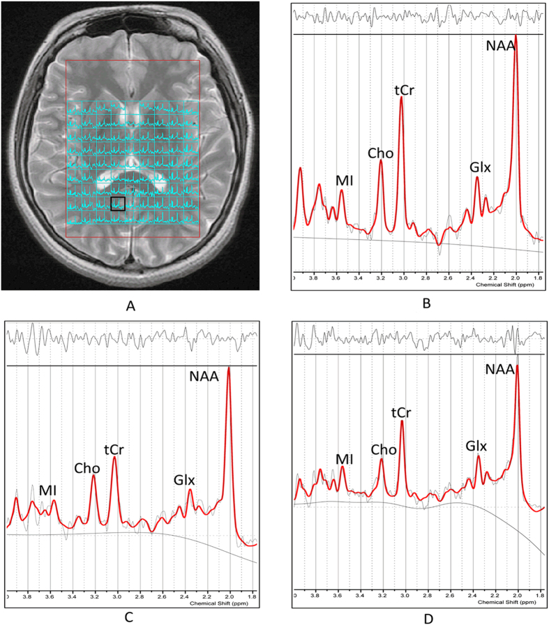
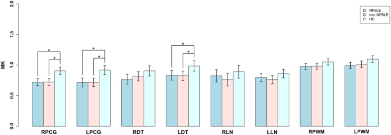
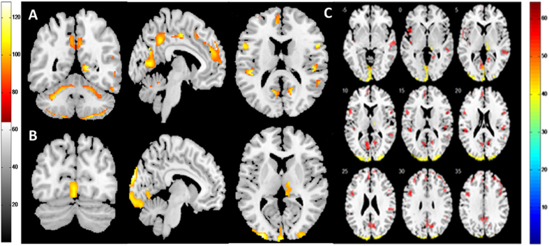
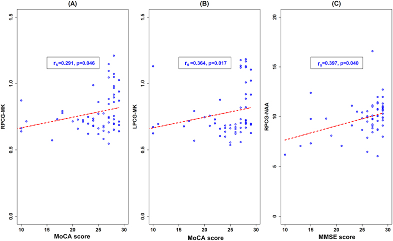
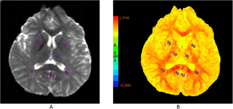
The diagnosis and pathology of neuropsychiatric systemic lupus erythematosus (NPSLE) remains challenging. Herein, we used multimodal imaging to assess anatomical and functional changes in brains of SLE patients instead of a single MRI approach generally used in previous studies. Twenty-two NPSLE patients, 21 non-NPSLE patients and 20 healthy controls (HCs) underwent 3.0 T MRI with multivoxel magnetic resonance spectroscopy, T1-weighted volumetric images for voxel based morphometry (VBM) and diffusional kurtosis imaging (DKI) scans. While there were findings in other basal ganglia regions, the most consistent findings were observed in the posterior cingulate gyrus (PCG). The reduction of multiple metabolite concentration was observed in the PCG in the two patient groups, and the NPSLE patients were more prominent. The two patient groups displayed lower diffusional kurtosis (MK) values in the bilateral PCG compared with HCs (p < 0.01) as assessed by DKI. Grey matter reduction in the PCG was observed in the NPSLE group using VBM. Positive correlations among cognitive function scores and imaging metrics in bilateral PCG were detected. Multimodal imaging is useful for evaluating SLE subjects and potentially determining disease pathology. Impairments of cognitive function in SLE patients may be interpreted by metabolic and microstructural changes in the PCG.
Figures
Similar articles
-
A multimodal MRI approach to identify and characterize microstructural brain changes in neuropsychiatric systemic lupus erythematosus.Neuroimage Clin. 2015 May 16;8:337-44. doi: 10.1016/j.nicl.2015.05.002. eCollection 2015. Neuroimage Clin. 2015. PMID: 26106559 Free PMC article.
-
Glial and axonal changes in systemic lupus erythematosus measured with diffusion of intracellular metabolites.Brain. 2016 May;139(Pt 5):1447-57. doi: 10.1093/brain/aww031. Epub 2016 Mar 11. Brain. 2016. PMID: 26969685 Free PMC article.
-
Brain histopathology in patients with systemic lupus erythematosus: identification of lesions associated with clinical neuropsychiatric lupus syndromes and the role of complement.Rheumatology (Oxford). 2017 Jan;56(1):77-86. doi: 10.1093/rheumatology/kew341. Epub 2016 Oct 25. Rheumatology (Oxford). 2017. PMID: 28028157
-
Quantitative magnetic resonance imaging in neuropsychiatric systemic lupus erythematosus.Lupus. 2003;12(12):897-902. doi: 10.1191/0961203303lu499oa. Lupus. 2003. PMID: 14714908 Review.
-
Neuropsychiatric lupus: clinical and imaging aspects.Bull NYU Hosp Jt Dis. 2007;65(3):194-9. Bull NYU Hosp Jt Dis. 2007. PMID: 17922669 Review.
Cited by
-
Diffusion Kurtosis Imaging of Microstructural Alterations in the Brains of Paediatric Patients with Congenital Sensorineural Hearing Loss.Sci Rep. 2017 May 8;7(1):1543. doi: 10.1038/s41598-017-01263-9. Sci Rep. 2017. PMID: 28484279 Free PMC article.
-
Laboratory and Neuroimaging Biomarkers in Neuropsychiatric Systemic Lupus Erythematosus: Where Do We Stand, Where To Go?Front Med (Lausanne). 2018 Dec 4;5:340. doi: 10.3389/fmed.2018.00340. eCollection 2018. Front Med (Lausanne). 2018. PMID: 30564579 Free PMC article. Review.
-
Cognitive Impairment in SLE: Mechanisms and Therapeutic Approaches.Curr Rheumatol Rep. 2021 Mar 29;23(4):25. doi: 10.1007/s11926-021-00992-1. Curr Rheumatol Rep. 2021. PMID: 33782842 Free PMC article. Review.
-
Quantitative morphometric changes in vascular mild cognitive impairment patients: early diagnosis of dementia.Cereb Cortex. 2023 Apr 25;33(9):5501-5506. doi: 10.1093/cercor/bhac437. Cereb Cortex. 2023. PMID: 36635220 Free PMC article.
-
Impaired decision-making and functional neuronal network activity in systemic lupus erythematosus.J Magn Reson Imaging. 2018 Dec;48(6):1508-1517. doi: 10.1002/jmri.26006. Epub 2018 Mar 14. J Magn Reson Imaging. 2018. PMID: 29537670 Free PMC article.
References
-
- Hochberg M. C. Updating the American College of Rheumatology revised criteria for the classification of systemic lupus erythematosus. Arthritis Rheum. 40, 1725–1725 (1997). - PubMed
-
- Adelmann D. C., Saltiel E. & Klinenberg J. R. The neuropsychiatric manifestations of SLE: an overview. Semin Arthritis Rheum. 15, 185–199 (1986). - PubMed
-
- Brey R. L. Neuropsychiatric lupus: clinical and imaging aspects. Bull NYU Hosp Jt Dis. 65, 194–199 (2007). - PubMed
-
- Toledano P., Sarbu N., Espinosa G., Bargallo N. & Cervera R. Neuropsychiatric systemic lupus erythematosus: magnetic resonance imaging findings and correlation with clinical and immunological features. Autoimmun Rev. 12, 1166–1170 (2013). - PubMed
-
- Sarbu N. et al. Brain abnormalities in newly diagnosed neuropsychiatric lupus: systematic MRI approach and correlation with clinical and laboratory data in a large multicenter cohort. Autoimmun Rev. 14, 153–159 (2015). - PubMed
Publication types
MeSH terms
LinkOut - more resources
Full Text Sources
Other Literature Sources
Medical
Molecular Biology Databases
