. 2016 Jan 12;7(1):e02001-15.
doi: 10.1128/mBio.02001-15.

Molecular and Structural Analysis of the Helicobacter pylori cag Type IV Secretion System Core Complex

Affiliations

Molecular and Structural Analysis of the Helicobacter pylori cag Type IV Secretion System Core Complex

Arwen E Frick-Cheng et al. mBio. .

Abstract

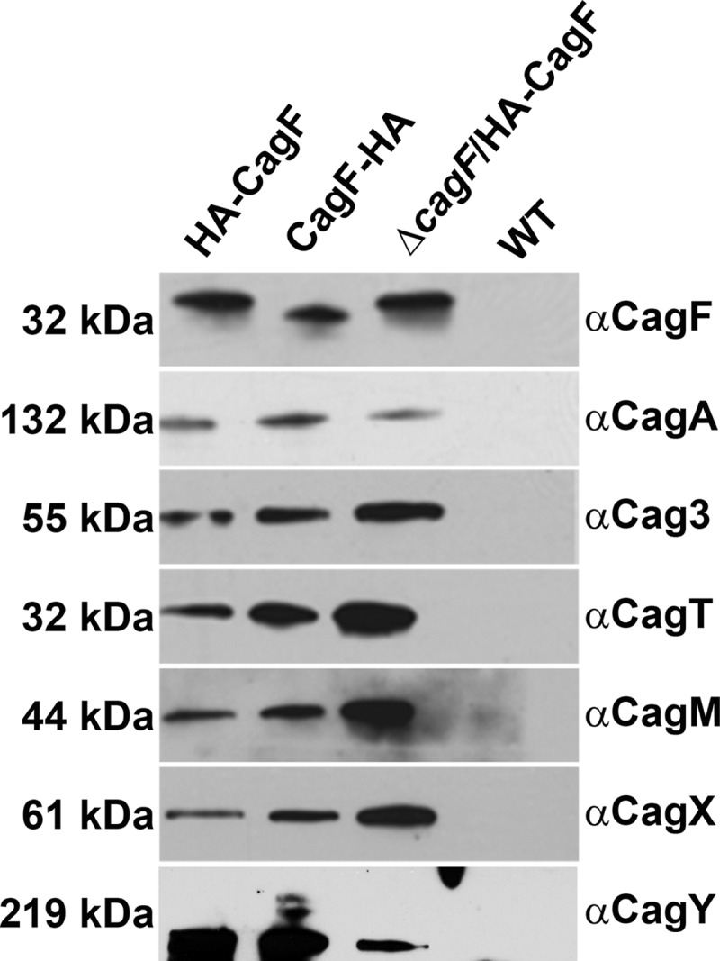
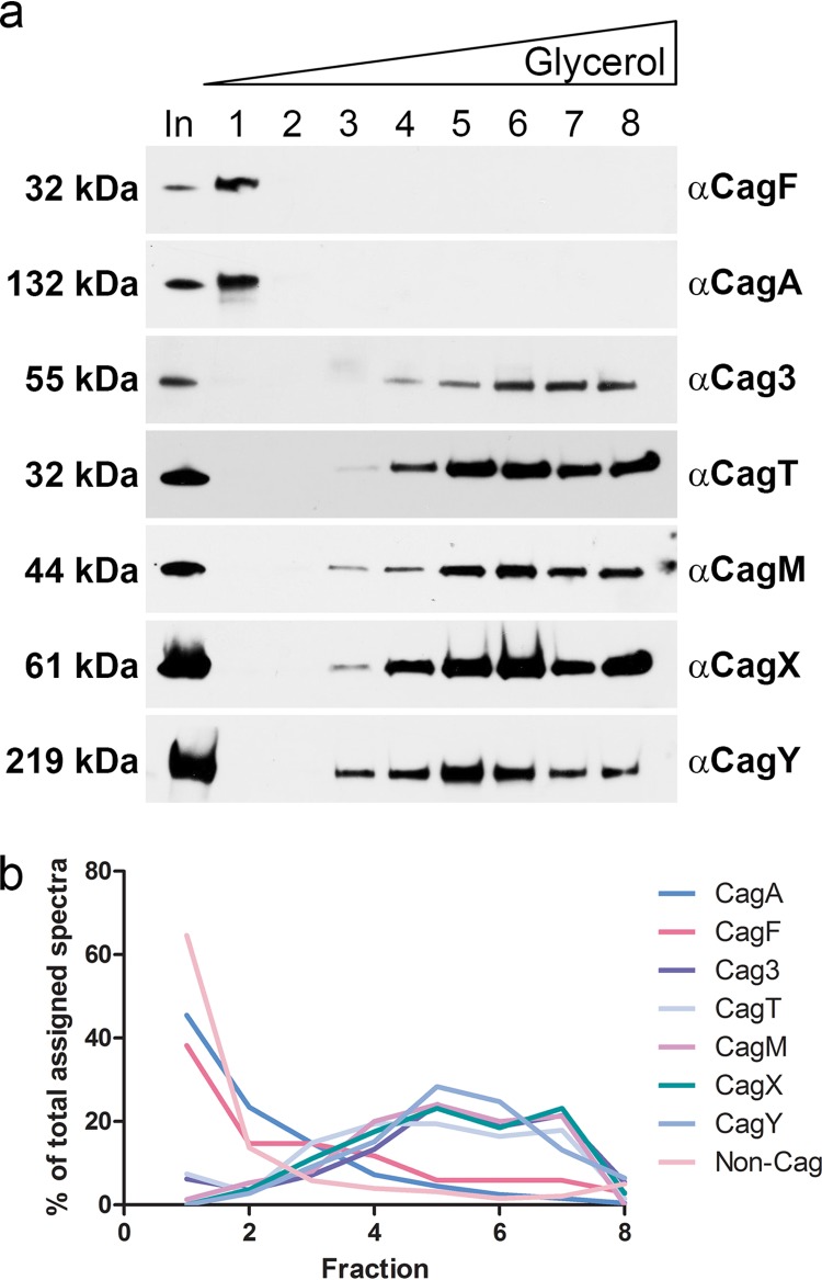
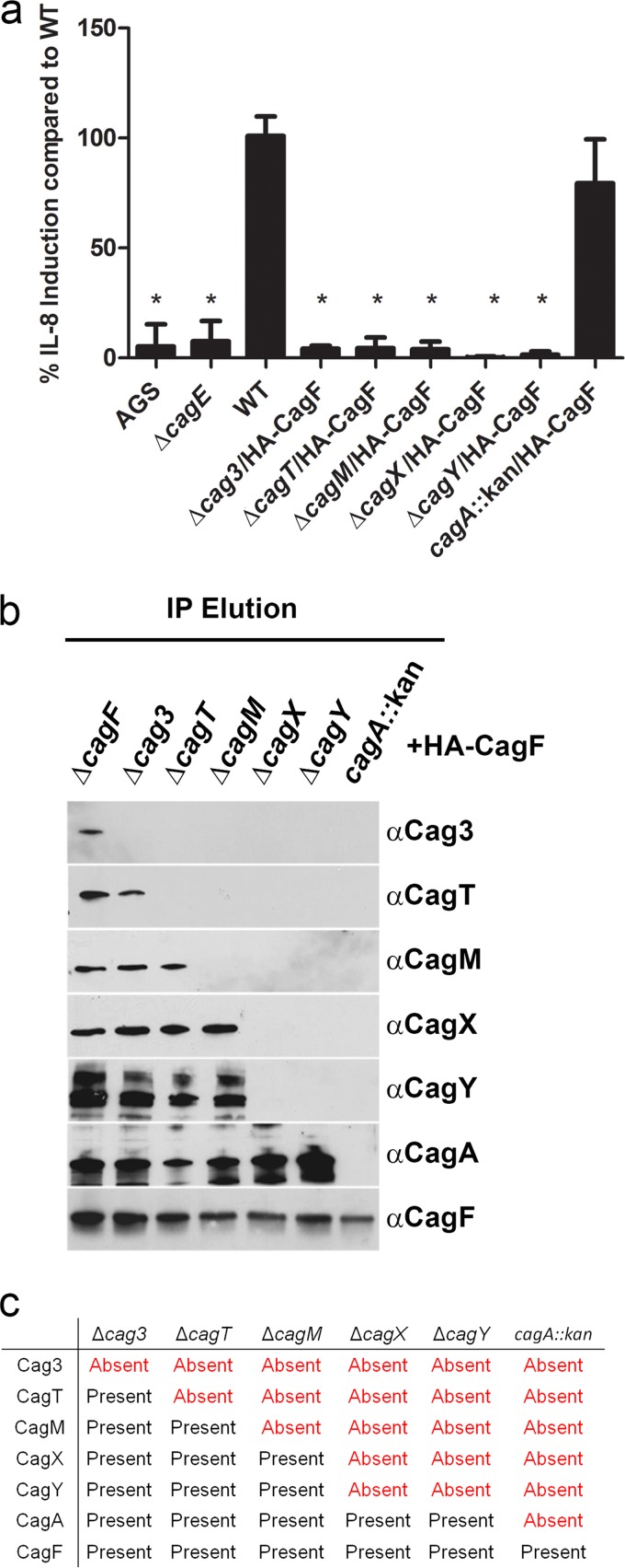
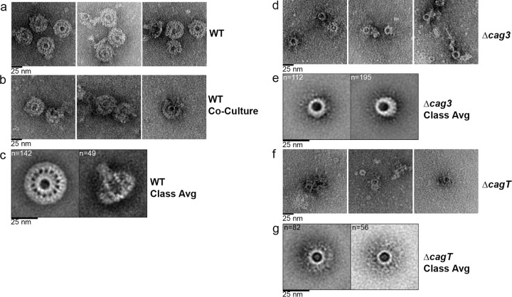
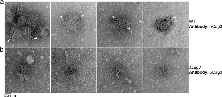
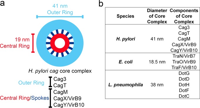
Bacterial type IV secretion systems (T4SSs) can function to export or import DNA, and can deliver effector proteins into a wide range of target cells. Relatively little is known about the structural organization of T4SSs that secrete effector proteins. In this report, we describe the isolation and analysis of a membrane-spanning core complex from the Helicobacter pylori cag T4SS, which has an important role in the pathogenesis of gastric cancer. We show that this complex contains five H. pylori proteins, CagM, CagT, Cag3, CagX, and CagY, each of which is required for cag T4SS activity. CagX and CagY are orthologous to the VirB9 and VirB10 components of T4SSs in other bacterial species, and the other three Cag proteins are unique to H. pylori. Negative stain single-particle electron microscopy revealed complexes 41 nm in diameter, characterized by a 19-nm-diameter central ring linked to an outer ring by spoke-like linkers. Incomplete complexes formed by Δcag3 or ΔcagT mutants retain the 19-nm-diameter ring but lack an organized outer ring. Immunogold labeling studies confirm that Cag3 is a peripheral component of the complex. The cag T4SS core complex has an overall diameter and structural organization that differ considerably from the corresponding features of conjugative T4SSs. These results highlight specialized features of the H. pylori cag T4SS that are optimized for function in the human gastric mucosal environment.

Importance: Type IV secretion systems (T4SSs) are versatile macromolecular machines that are present in many bacterial species. In this study, we investigated a T4SS found in the bacterium Helicobacter pylori. H. pylori is an important cause of stomach cancer, and the H. pylori T4SS contributes to cancer pathogenesis by mediating entry of CagA (an effector protein regarded as a "bacterial oncoprotein") into gastric epithelial cells. We isolated and analyzed the membrane-spanning core complex of the H. pylori T4SS and showed that it contains unique proteins unrelated to components of T4SSs in other bacterial species. These results constitute the first structural analysis of the core complex from this important secretion system.


Figures

FIG 1
FIG 1
Analysis of proteins that copurify with CagF. Immunoblot analysis of preparations immunopurified from three strains producing HA-tagged CagF using a monoclonal HA antibody and of a control preparation from an untagged WT strain that was processed in parallel. Samples were immunoblotted with the indicated antisera. Results are representative of at least two independent experiments.
FIG 2
FIG 2
Sedimentation of H. pylori cag T4SS core complex in density gradients. (a) Preparations resulting from immunoaffinity purification of HA-CagF were analyzed by velocity sedimentation in glycerol density gradients, and gradient fractions (collected from the top of the gradient) were immunoblotted with the indicated antisera. In, input sample before it was applied to the gradient. Results are representative of two independent experiments. (b) The protein content of gradient fractions was analyzed by 1D mass spectrometry (see Table S3 in the supplemental material for complete results). The distribution of CagF, CagA, Cag3, CagT, CagM, CagX, CagY, and total non-Cag proteins in individual gradient fractions is shown.
FIG 3
FIG 3
Immunopurification of HA-CagF from a panel of cag mutant strains. (a) AGS cells were cocultured with the WT strain or the indicated mutants, and IL-8 secretion was quantified by enzyme-linked immunosorbent assay (ELISA). “AGS” indicates cells without added H. pylori. Values represent means ± standard deviations (SD), based on analysis of at least six replicate samples. Levels of IL-8 production induced by mutant strains were compared to levels induced by the WT strain (analysis of variance [ANOVA] followed by Dunn’s multiple-comparison test). *, P < 0.001. (b) HA-CagF was immunoaffinity purified from the strains analyzed in panel A. The affinity-purified samples (immunopurification [IP] elution) were then immunoblotted with the indicated antisera. Strains are designated at the top of the panel, and antisera are designated at the right side of the panel. (c) Summary of results from the experiments presented in panel B, indicating the presence or absence of Cag proteins in the preparations immunopurified from mutant strains. Results are representative of three independent experiments.
FIG 4
FIG 4
Negative-stain EM analysis of the H. pylori cag T4SS core complex. (a) Representative negative-stain images of WT cag T4SS complexes purified from a strain with an intact cag PAI that was engineered to produce HA-CagF. (b) Representative negative-stain images of WT cag T4SS complexes purified from an HA-CagF-producing strain (ΔcagF/HA-CagF) cocultured with AGS cells. (c) Class averages (Class Avg) showing an en face view and a side view of WT complexes. (d) Representative negative-stain images of cag T4SS complexes purified from a Δcag3 strain engineered to produce HA-CagF. (e) Class averages of the cag T4SS complex purified from a Δcag3 strain. (f) Representative negative-stain images of cag T4SS complexes purified from a ΔcagT strain engineered to produce HA-CagF. (g) Class averages of the cag T4SS complex purified from a ΔcagT strain. Numbers of particles used for generating each average are shown in upper left corner of panels. All scale bars represent 25 nm.
FIG 5
FIG 5
Localization of Cag3 by immunogold labeling and negative-stain EM. Negative-stain EM images of WT core complexes, isolated from a strain with an intact cag PAI (a) or a Δcag3 mutant (b) and subjected to immunogold labeling with primary anti-Cag3 antiserum followed by 5-nm-diameter-gold-particle-conjugated secondary antibodies, are shown. Arrowheads indicate representative gold particles. Images are representative of results from three independent experiments. Scale bar, 25 nm.
FIG 6
FIG 6
Schematic model of the H. pylori cag T4SS core complex. (a) Prominent features of the complex include an outer ring (light blue) and central ring (red) connected by 14 spokes (dark blue). Analysis of complexes from mutant strains suggests that Cag3 and CagT are localized to the periphery of the intact wild-type complex. (b) Comparison of the diameters and components of T4SS core complexes from H. pylori, E. coli, and L. pneumophila.

References

    1. Robin Warren J, Marshall B. 1983. Unidentified curved bacilli on gastric epithelium in active chronic gastritis. Lancet 321:1273–1275. doi:10.1016/S0140-6736(83)92719-8. - DOI - PubMed
    1. Cover TL, Blaser MJ. 2009. Helicobacter pylori in health and disease. Gastroenterology 136:1863–1873. doi:10.1053/j.gastro.2009.01.073. - DOI - PMC - PubMed
    1. Peek RM Jr, Blaser MJ. 2002. Helicobacter pylori and gastrointestinal tract adenocarcinomas. Nat Rev Cancer 2:28–37. doi:10.1038/nrc703. - DOI - PubMed
    1. Atherton JC. 2006. The pathogenesis of Helicobacter pylori-induced gastroduodenal diseases. Annu Rev Pathol 1:63–96. doi:10.1146/annurev.pathol.1.110304.100125. - DOI - PubMed
    1. Suerbaum S, Michetti P. 2002. Helicobacter pylori infection. N Engl J Med 347:1175–1186. doi:10.1056/NEJMra020542. - DOI - PubMed

Publication types

MeSH terms

Substances