Review
. 2016 Feb 1;35(3):239-57.
doi: 10.15252/embj.201592705. Epub 2016 Jan 12.

Astrocytes as secretory cells of the central nervous system: idiosyncrasies of vesicular secretion

Affiliations
Review

Astrocytes as secretory cells of the central nervous system: idiosyncrasies of vesicular secretion

Alexei Verkhratsky et al. EMBO J. .

Abstract

Astrocytes are housekeepers of the central nervous system (CNS) and are important for CNS development, homeostasis and defence. They communicate with neurones and other glial cells through the release of signalling molecules. Astrocytes secrete a wide array of classic neurotransmitters, neuromodulators and hormones, as well as metabolic, trophic and plastic factors, all of which contribute to the gliocrine system. The release of neuroactive substances from astrocytes occurs through several distinct pathways that include diffusion through plasmalemmal channels, translocation by multiple transporters and regulated exocytosis. As in other eukaryotic cells, exocytotic secretion from astrocytes involves divergent secretory organelles (synaptic-like microvesicles, dense-core vesicles, lysosomes, exosomes and ectosomes), which differ in size, origin, cargo, membrane composition, dynamics and functions. In this review, we summarize the features and functions of secretory organelles in astrocytes. We focus on the biogenesis and trafficking of secretory organelles and on the regulation of the exocytotic secretory system in the context of healthy and diseased astrocytes.

Keywords: SNARE proteins; astrocytes; exocytosis; secretion; secretory vesicles.


Figures

Figure 1
Figure 1. Multiple secretory pathways operating in astrocyte
Figure 2
Figure 2. Arrangement of VAMP2 on vesicles in astrocytes
(A) The diagram represents an astrocytic vesicle with the architecture of VAMP2 across the vesicle membrane. VAMP2 is appended at its luminal C‐terminus with yellow phluorin (YpH, shown in green) and can be immunolabelled at its N‐terminus (cytoplasmic site) using primary and secondary antibodies, the latter tagged with Atto594, a red fluorophore. The graph (below) shows the measurements of distance between the two fluorophores obtained from an astrocyte shown in (B), indicating the length of VAMP2 in astrocytes. (B) Structured illumination microscopy (SIM) micrograph of an astrocyte expressing VAMP2 marked fluorescently at luminal and cytoplasmic sites as schematized in (A). There is an incomplete co‐localisation, that is separation, between the red and green puncta, disclosed as the distance in (A); co‐localisation of YpH and Atto594 is disclosed in yellow. An area (box) of an astrocyte is shown in the inset (bottom right). Scale bar: 10 µm (300 nm in inset). (Modified with permission from Singh et al, 2014).
Figure 3
Figure 3. Diversity of astroglial vesicles
Bottom panel shows schematic overview of astroglial secretory organelles. Top panel demonstrates ultrastructure of astroglial secretory organelles. (a) Electron micrograph of small clear vesicles (red arrowheads) in an astrocyte (Ast) from the rat hippocampus in situ. Scale bars: 100 nm (50 nm in inset). Modified with permission from Bergersen et al (2012). (b) Small clear vesicles from cultured astrocytes. Courtesy of Dr. Michela Matteoli. Scale bar: 50 nm. (c) Electron micrograph of a dense‐core vesicle in cultured astrocytes stained by immunogold for secretogranin II. Modified with permission from Calegari et al (1999). Arrow shows DCV stained for secretogranin II. The open arrowhead points to the intermediate filament immunolabelled for GFAP. Scale bar: 100 nm. (d) Electron micrograph showing a dense‐core vesicle in cultured astrocytes. Scale bar: 500 nm. Modified with permission from Prada et al (2011). (e) Electron micrograph of lysosomes (L) in astrocytes. Scale bar: 1 µm. ©2012 by National Academy of Sciences. Modified with permission from Di Malta et al (2012). (f) Electron microscopy images of an ADF glioma cell. Arrows point to multilamellar organelles. Scale bar: 150 nm. Modified with permission from Verderio et al (2012). (g) Electron micrograph showing a multivesicular body‐like structure from a rat cultured astrocyte. Arrowheads indicate vesicles. Scale bars: 200 µm. Modified with permission from Brignone et al (2015). (h) Electron micrograph (negative staining) showing an exosome secreted by cultured astrocytes following stimulation with 100 mM BzATP, a P2X agonist, for 20 min. Scale bar: 300 nm. Modified with permission from Bianco et al (2009). Abbreviations: aSMase, acid sphingomyelinase; BMP, bis(monoacylglycero)phosphate; FGF2, fibroblast growth factor 2; Hsp70, 70 kilodalton heat shock protein; miRNA, microRNA; MMPs, matrix metalloproteinases; mtRNA, mitochondrial RNA; nSMase2, neutral sphingomyelinase 2; NTPDase, nucleoside triphosphate diphosphohydrolases; PS, phosphatidylserine; REST, RE‐1‐silencing transcription factor; tPA, tissue plasminogen activator; VGEF, vascular endothelial growth factor.
Figure 4
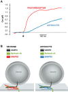
Figure 4. Slowness of astroglial exocytosis
(A) Comparison of kinetics of neuronal and astroglial exocytosis. Time‐dependent changes in membrane capacitance (Cm) recorded in a neuronal cell (trace in red, photoreceptor, modified from Kreft et al, 2003) and an astrocyte (trace in blue, modified from Kreft et al, 2004), elicited by a flash photolysis‐induced increase in cytosolic Ca2+. Note that the blue trace recorded in an astrocyte displays a significant delay between the stimulus (asterisk) and the response (trace components above the dotted line). (B) Neuronal vs. astrocytic SNAREs. Neurones and astrocytes alike express SNAREs VAMP2 and syntaxin 1; many astrocytes can also express VAMP3 in lieu of or in addition to VAMP2. Astrocytes express SNAP23, a homologue of neuronal SNAP25. At the plasma membrane, syntaxin 1A can form a binary cis complex with SNAP25B or SNAP23A, which then interacts with vesicular VAMP2 to form a ternary complex. A single ternary complex can tether the vesicle at the plasma membrane for a longer period of time (1.9 s vs. 0.8 s), when it contains SNAP25B rather than SNAP23A, respectively. Of note, truncated syntaxin 1, lacking the N‐terminal Habc domain and the linker region to the SNARE domain, is shown for simplicity. Drawings are not to scale.

References

    1. Andrews NW, Chakrabarti S (2005) There's more to life than neurotransmission: the regulation of exocytosis by synaptotagmin VII. Trends Cell Biol 15: 626–631 - PubMed
    1. Arcienega II, Brunet JF, Bloch J, Badaut J (2010) Cell locations for AQP1, AQP4 and 9 in the non‐human primate brain. Neuroscience 167: 1103–1114 - PubMed
    1. Arima H, Yamamoto N, Sobue K, Umenishi F, Tada T, Katsuya H, Asai K (2003) Hyperosmolar mannitol simulates expression of aquaporins 4 and 9 through a p38 mitogen‐activated protein kinase‐dependent pathway in rat astrocytes. J Biol Chem 278: 44525–44534 - PubMed
    1. Baietti MF, Zhang Z, Mortier E, Melchior A, Degeest G, Geeraerts A, Ivarsson Y, Depoortere F, Coomans C, Vermeiren E, Zimmermann P, David G (2012) Syndecan‐syntenin‐ALIX regulates the biogenesis of exosomes. Nat Cell Biol 14: 677–685 - PubMed
    1. Barg S, Ma X, Eliasson L, Galvanovskis J, Gopel SO, Obermuller S, Platzer J, Renstrom E, Trus M, Atlas D, Striessnig J, Rorsman P (2001) Fast exocytosis with few Ca2+ channels in insulin‐secreting mouse pancreatic β cells. Biophys J 81: 3308–3323 - PMC - PubMed

Publication types

Substances