. 2016 Jan 13;36(2):405-18.
doi: 10.1523/JNEUROSCI.2555-15.2016.

Isolated P/Q Calcium Channel Deletion in Layer VI Corticothalamic Neurons Generates Absence Epilepsy

Affiliations

Isolated P/Q Calcium Channel Deletion in Layer VI Corticothalamic Neurons Generates Absence Epilepsy

Valerie C Bomben et al. J Neurosci. .

Abstract

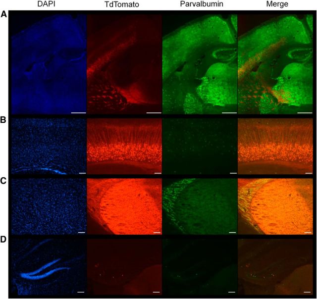
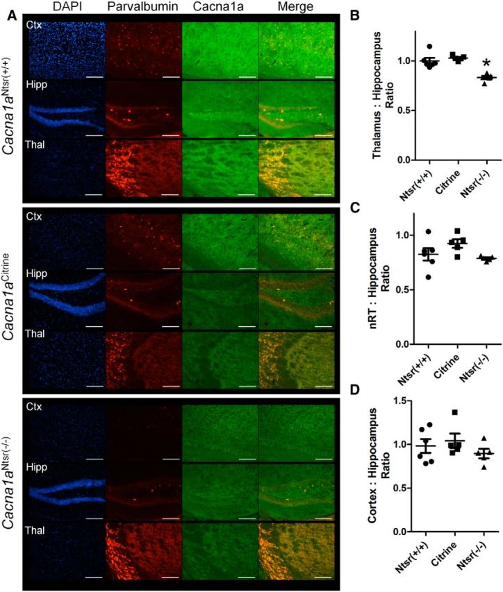
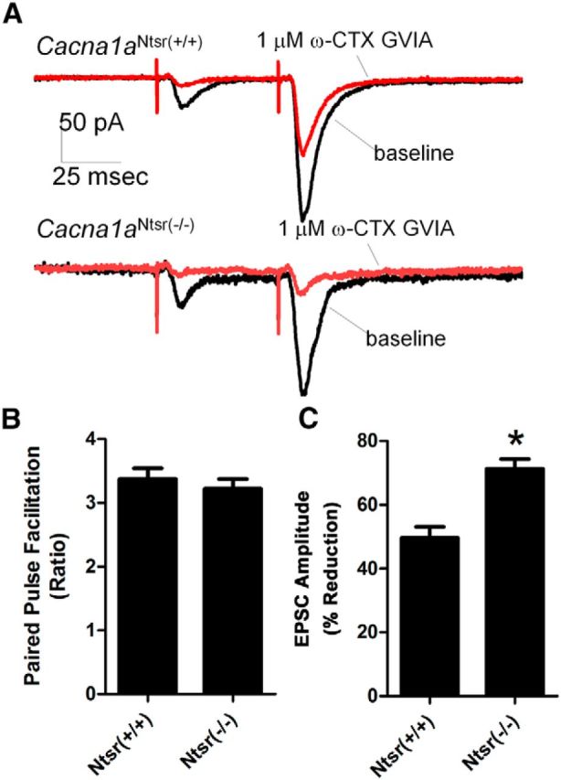
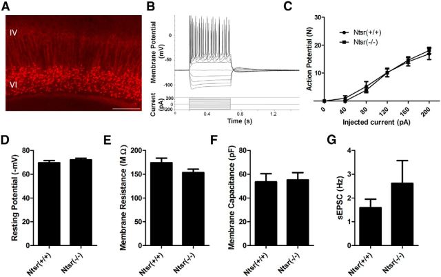
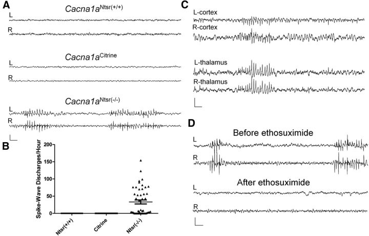
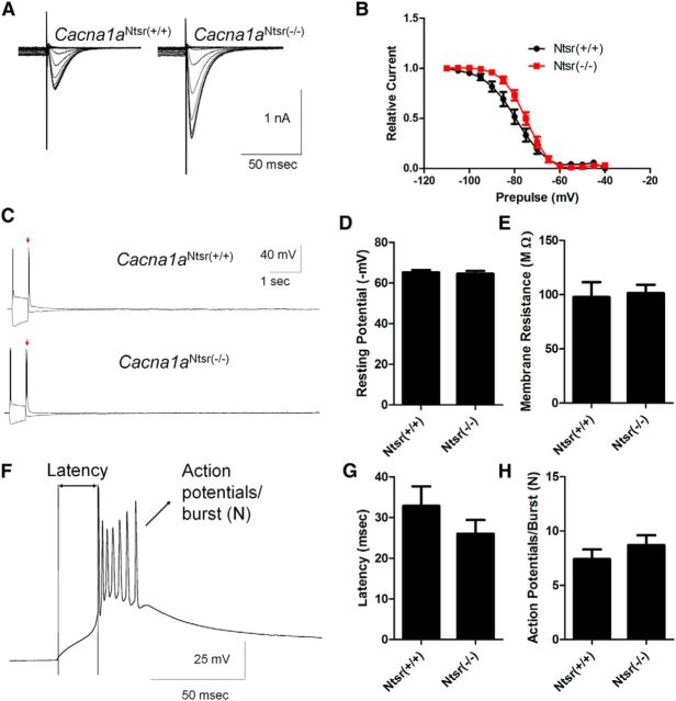
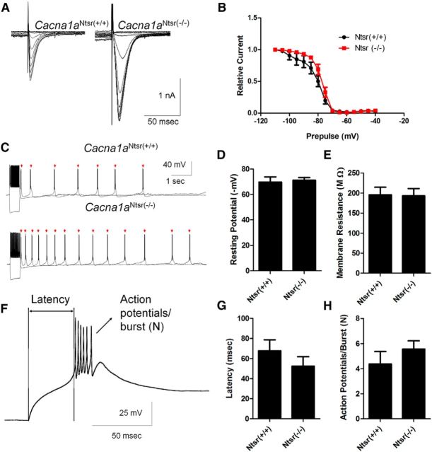
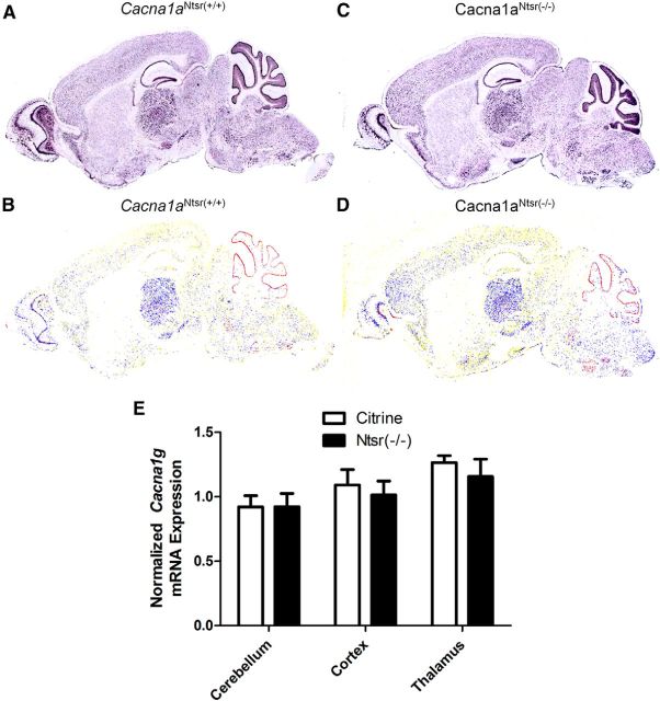
Generalized spike-wave seizures involving abnormal synchronization of cortical and underlying thalamic circuitry represent a major category of childhood epilepsy. Inborn errors of Cacna1a, the P/Q-type voltage-gated calcium channel α subunit gene, expressed throughout the brain destabilize corticothalamic rhythmicity and produce this phenotype. To determine the minimal cellular lesion required for this network disturbance, we used neurotensin receptor 1 (Ntsr1) cre-driver mice to ablate floxed Cacna1a in layer VI pyramidal neurons, which supply the sole descending cortical synaptic input to thalamocortical relay cells and reticular interneurons and activate intrathalamic circuits. Targeted Cacna1a ablation in layer VI cells resulted in mice that display a robust spontaneous spike-wave absence seizure phenotype accompanied by behavioral arrest and inhibited by ethosuximide. To verify the selectivity of the molecular lesion, we determined that P/Q subunit proteins were reduced in corticothalamic relay neuron terminal zones, and confirmed that P/Q-mediated glutamate release was reduced at these synapses. Spike-triggered exocytosis was preserved by N-type calcium channel rescue, demonstrating that evoked release at layer VI terminals relies on both P/Q and N-type channels. Whereas intrinsic excitability of the P/Q channel depleted layer VI neurons was unaltered, T-type calcium currents in the postsynaptic thalamic relay and reticular cells were dramatically elevated, favoring rebound bursting and seizure generation. We find that an early P/Q-type release defect, limited to synapses of a single cell-type within the thalamocortical circuit, is sufficient to remodel synchronized firing behavior and produce a stable generalized epilepsy phenotype.

Significance statement: This study dissects a critical component of the corticothalamic circuit in spike-wave epilepsy and identifies the developmental importance of P/Q-type calcium channel-mediated presynaptic glutamate release at layer VI pyramidal neuron terminals. Genetic ablation of Cacna1a in layer VI neurons produced synchronous spike-wave discharges in the cortex and thalamus that were inhibited by ethosuximide. These mice also displayed N-type calcium channel compensation at descending thalamic synapses, and consistent with other spike-wave models increased low-threshold T-type calcium currents within postsynaptic thalamic relay and reticular neurons. These results demonstrate, for the first time, that preventing the developmental homeostatic switch from loose to tightly coupled synaptic release at a single class of deep layer cortical excitatory output neurons results in generalized spike-wave epilepsy.

Keywords: Cacna1a; T-type; mouse; plasticity; spike-wave; tottering.


Figures

Figure 1.
Figure 1.
The Ntsr-1 cre drives expression of tdTomato in layer VI projection neurons to the thalamic reticular nucleus and ventrobasal thalamus. A, Stitched 10× images of the projections from layer VI through the internal capsule to the nRT and thalamus. Cell nuclei are marked with DAPI in blue, TdTomato staining in red indicates Ntsr Cre+ cells, and FITC staining in green marks parvalbumin interneurons throughout the brain. Scale bar, 500 μm. B, Image (10×) of layer VI cortical projection neurons. Scale bar, 200 μm. C, Image (10×) of the nRT and thalamus shows no cre-driven cellular staining, but diffuse label due to axonal projections from layer VI neurons. Scale bar, 200 μm. D, Image (10×) of hippocampal dentate gyrus shows scattered cells expressing tdTomato. Scale bar, 200 μm.
Figure 2.
Figure 2.
Immunostaining reveals specific knockdown of Cacna1a in ventrobasal regions of the thalamus. A, Representative staining in (top) wild-type (Cacna1aNtsr(+/+), (middle) Cacna1aCitrine, and (lower) Cacna1aNtsr(−/−)mice of cortex, hippocampus, and thalamus. TRITC-conjugated parvalbumin staining was used to identify the nRT. FITC staining marks P/Q calcium channels and DAPI marks cell nuclei. Scale bars, 100 μm. B, Densitometric ratio of the ventrobasal thalamus compared with the mossy fiber region of the hippocampus within slices of wild-type (Cacna1aNtsr(+/+), n = 6, 1.00 ± 0.03; Cacna1aCitrine, n = 5, 1.03 ± 0.01; Cacna1aNtsr(−/−), n = 5, 0.83 ± 0.02) reveal decreased P/Q immunostaining (ANOVA, *p = 0.0001). C, Densitometric ratio of the nRT compared with the mossy fiber region of the hippocampus within slices of wild-type (Cacna1aNtsr(+/+), n = 6, 0.82 ± 0.06; Cacna1aCitrine, n = 5, 0.92 ± 0.04; Cacna1aNtsr(−/−), n = 5, 0.79 ± 0.09) show no decreased P/Q immunostaining (ANOVA, p = 0.11). D, Densitometric ratio of the layer V/VI cortex compared with the mossy fiber region of the hippocampus within slices of wild-type (Cacna1aNtsr(+/+), n = 6, 0.98 ± 0.08; Cacna1aCitrine, n = 5, 1.04 ± 0.08; Cacna1aNtsr(−/−), n = 5, 0.89 ± 0.05) show no decreased P/Q immunostaining (ANOVA, p = 0.50).
Figure 3.
Figure 3.
Electrophysiological recordings from the cortical thalamic synapse reveal functional loss of P/Q calcium channels. A, Example traces of PPF within the ventrobasal thalamus when stimulated in the internal capsule before and after the application of N-type calcium channel blocker, ω-CTX GVIA. Scale bar, 50 ms. B, Baseline PPF is not significantly different (t test, p = 0.51) between Cacna1aNtsr(+/+) (PPF ratio 3.4 ± 0.6, n = 12) and Cacna1aNtsr(−/−) mice (PPF ratio 3.2 ± 0.5, n = 12). C, Application of 1 μm ω-CTX GVIA resulted in a significant EPSC decrease (t test, *p = 0.0001) of 50 ± 12% in Cacna1aNtsr(+/+) slices (n = 12) and 71 ± 11% in Cacna1aNtsr(−/−) slices (n = 12).
Figure 4.
Figure 4.
Whole-cell patch-clamp electrophysiology of layer VI cells. A, Representative tdTomato expression in layer VI for identification to patch-clamp. Scale bar, 200 μm. B, Representative voltage step protocol and current response by a Cacna1aNtsr(+/+) cell. C, Number of action potentials elicited by injected positive current for Cacna1aNtsr(+/+) cells (n = 9) and Cacna1aNtsr(−/−) cells (n = 9). No significant difference was found at any current step. (ANOVA, p = 0.88 and 0.91 for column factor or interaction). D, No difference in resting membrane potential of layer VI projection neurons. The mean resting membrane potential for Cacna1aNtsr(+/+) cells was 69.5 ± 1.9 mV and for Cacna1aNtsr(−/−) cells 72.0 ± 1.4 mV (n = 9 each, t test, p = 0.30). E, No difference in membrane resistance of layer VI projection neurons. The mean membrane resistance for Cacna1aNtsr(+/+) cells was 174.0 ± 9.7 MΩ and for Cacna1aNtsr(−/−) cells 153.5 ± 7.0 MΩ (n = 9 each, t test, p = 0.11). F, No difference in membrane capacitance of layer VI projection neurons. The mean membrane capacitance for Cacna1aNtsr(+/+) cells was 53.5 ± 6.9 pF and for Cacna1aNtsr(−/−) cells was 55.2 ± 6.1 pF (n = 9 each, t test, p = 0.85). G, No difference in the frequency of spontaneous EPSCs of layer VI projection neurons. Frequency of spontaneous EPSCs for Cacna1aNtsr(+/+) cells was 1.59 ± 0.36 Hz and for Cacna1aNtsr(−/−) cells 2.61 ± 0.96 Hz (n = 9 each, t test with Welch's correction, p = 0.34).
Figure 5.
Figure 5.
Cacna1aNtsr(−/−) mice have spike-wave epilepsy. A, Representative 12 s traces of wild-type (Cacna1aNtsr(+/+), Cacna1aCitrine) and Cacna1aNtsr(−/−) mice electroencephalographic recordings from bilateral electrodes over frontal cortex. Cacna1aNtsr(−/−) mice display stereotyped 5–7 spike/s spike-wave discharges. Scale bar, 500 μV and 500 ms. B, Spike-wave discharge rates from Cacna1aNtsr(+/+), N = 3, 12 h/mouse; Cacna1aCitrine, N = 4, 12 h/mouse; and Cacna1aNtsr(−/−) mice, N = 4, 12 h/mouse. Spike-wave discharges were only seen in Cacna1aNtsr(−/−) mice. C, Representative bilateral EEG recording from the frontal cortex with depth electrodes placed in the ventrobasal thalamus show spike-wave discharges in both regions of the brain. Scale bar, 500 μV and 500 ms. D, Representative EEG recording from a Cacna1aNtsr(−/−) frontal cortex before and after intraperitoneal injection of 200 mg/kg ethosuximide shows suppression of spike-wave discharges after drug exposure. Scale bar, 500 μV and 500 ms.
Figure 6.
Figure 6.
T-type calcium channel currents are increased in Cacna1aNtsr(−/−) thalamic relay neurons. A, Sample traces of low voltage-activated calcium currents from ventrobasal thalamic relay neurons in Cacna1aNtsr(+/+) and Cacna1aNtsr(−/−) cells with prior hyperpolarization steps to activate the currents. Currents were elevated 1.7-fold in Cacna1aNtsr(−/−) cells. B, SSI (half-maximum voltage of the prior hyperpolarization voltage), showed a significant depolarizing shift in Cacna1aNtsr(−/−) ventrobasal thalamic neurons (−75.3 ± 1.2 mV, n = 10) from Cacna1aNtsr(+/+) (−82.2 ± 2.1 mV; t test, p = 0.011). C, Sample traces of rebound bursts from ventrobasal thalamic relay neurons in Cacna1aNtsr(+/+) and Cacna1aNtsr(−/−). No difference in rebound bursts was observed. D, Resting membrane potential of Cacna1aNtsr(+/+) (65.1 ± 1.2 mV, n = 7) and Cacna1aNtsr(−/−) (64.5 ± 1.4 mV, n = 7; t test, p = 0.80) cells. E, Membrane resistance of Cacna1aNtsr(+/+) (97.7 ± 13.7 MΩ, n = 7) and Cacna1aNtsr(−/−) (101.4 ± 7.6 MΩ, n = 7; t test, p = 0.18) cells. F, Representative trace of first rebound burst following hyperpolarization. Scale bar, 25 mV/50 ms. G, Latency to burst of Cacna1aNtsr(+/+) (32.9 ± 4.8 ms, n = 7) and Cacna1aNtsr(−/−) (26.0 ± 3.4 ms; n = 7; t test, p = 0.41) cells. H, Number (N) of action potentials in burst for Cacna1aNtsr(+/+) (7.4 ± 0.9 AP, n = 7) and Cacna1aNtsr(−/−) (8.7 ± 0.9 AP; n = 7; t test, p = 0.99) cells.
Figure 7.
Figure 7.
T-type calcium channel currents are increased in Cacna1aNtsr(−/−) reticular thalamic interneurons. A, Sample traces of low-voltage activated calcium currents from nRT neurons in Cacna1aNtsr(+/+) and Cacna1aNtsr(−/−) cells with prior hyperpolarization steps to activate the currents. B, SSI was not changed between in Cacna1aNtsr(+/+) (−81.6 ± 2.5 mV, n = 10) and Cacna1aNtsr(−/−) nRT neurons (−78.1 ± 1.0 mV, n = 10, t test with Welch's correction, p = 0.23). C, Sample traces of rebound bursts show a significant difference from nRT neurons in Cacna1aNtsr(+/+) (2.12 ± 0.79 bursts, n = 10) and Cacna1aNtsr(−/−) (10.57 ± 2.22 bursts, n = 10, t test, p = 0.009). D, Resting membrane potential of Cacna1aNtsr(+/+) (69.7 ± 4.1 mV, n = 8) and Cacna1aNtsr(−/−) (71.1 ± 2.2 mV, n = 7; t test, p = 0.10) cells. E, Membrane resistance of Cacna1aNtsr(+/+) (195.7 ± 19.1 MΩ, n = 8) and Cacna1aNtsr(−/−) (193.1 ± 18.2 MΩ, n = 7; t test, p = 0.79) cells. F, Representative trace of first rebound burst following hyperpolarization. Scale bar, 25 mV/50 ms. G, Latency to burst of Cacna1aNtsr(+/+) (67.8 ± 10.9 ms, n = 7) and Cacna1aNtsr(−/−) (52.5 ± 9.4 ms; n = 7; t test, p = 0.73) cells. H, Number of action potentials in burst for Cacna1aNtsr(+/+) (5.0 ± 0.9 AP, n = 7) and Cacna1aNtsr(−/−) (5.6 ± 0.6 AP; n = 7; t test, p = 0.41) cells.
Figure 8.
Figure 8.
Cacna1g mRNA transcript levels are not altered in Cacna1aNtsr(−/−) mice. A, RNA in situ hybridization of Cacna1g mRNA in Cacna1aNtsr(+/+) brain. B, RNA in situ hybridization of Cacna1g in Cacna1aNtsr(−/−) brain. C, Pseudocolored cell-based densitometry of in situ hybridization image of Cacna1g mRNA in Cacna1aNtsr(+/+) brain. D, Pseudocolored RNA in situ hybridization image of Cacna1g in Cacna1aNtsr(−/−) brain. E, Normalized ratio of Cacna1g mRNA transcripts revealed no difference in mRNA expression within either the cerebellum, the cortex, or the thalamus of Cacna1aNtsr(−/−) mice (n = 3) compared with Cacna1aNtsr(+/+) mice (black square, n = 3) or Cacna1aCitrine mice (red circle, n = 3).
Figure 9.
Figure 9.
Minimal motor deficit in Cacna1aNtsr(−/−) mutants. A, Rotarod: average latency to fall over three trials in wild-type (Cacna1aNtsr(+/+), n = 9, 111 ± 10 s; Cacna1aCitrine, n = 6, 101 ± 13 s; and Cacna1aNtsr(−/−), n = 9, 111 ± 16 s). No significant difference was observed (ANOVA, p = 0.89). B, Gait test: functional score (range 0–3) of wild-type (Cacna1aNtsr(+/+), n = 9, 0 ± 0; Cacna1aCitrine, n = 6, 0.17 ± 0.17; and Cacna1aNtsr(−/−), n = 9, 0.33 ± 0.17). No significant difference was observed (ANOVA, p = 0.17). C, Ledge test: functional score (range 0–3) of wild-type (Cacna1aNtsr(+/+), n = 11, 0.18 ± 0.12; Cacna1aCitrine, n = 11, 0.27 ± 0.14; and Cacna1aNtsr(−/−), n = 13, 0.85 ± 0.19, ANOVA, *p = 0.017), which was a small, but significant, deficit in motor coordination.

Similar articles

Cited by

References

    1. Agmon A, Connors BW. Thalamocortical responses of mouse somatosensory (barrel) cortex in vitro. Neuroscience. 1991;41:365–379. doi: 10.1016/0306-4522(91)90333-J. - DOI - PubMed
    1. Ali AB, Nelson C. Distinct Ca2+ channels mediate transmitter release at excitatory synapses displaying different dynamic properties in rat neocortex. Cereb Cortex. 2006;16:386–393. doi: 10.1093/cercor/bhi117. - DOI - PubMed
    1. Bal T, Debay D, Destexhe A. Cortical feedback controls the frequency and synchrony of oscillations in the visual thalamus. J Neurosci. 2000;20:7478–7488. - PMC - PubMed
    1. Baur D, Bornschein G, Althof D, Watanabe M, Kulik A, Eilers J, Schmidt H. Developmental tightening of cerebellar cortical synaptic influx-release coupling. J Neurosci. 2015;35:1858–1871. doi: 10.1523/JNEUROSCI.2900-14.2015. - DOI - PMC - PubMed
    1. Berger H. Über das elektrenkephalogramm des menschen. Archiv Psychiatri Nervenkrankheit. 1933;98:231–254. doi: 10.1007/BF01814645. - DOI

Publication types

MeSH terms

LinkOut - more resources