Clinical Trial
. 2016 Jan 13:16:5.
doi: 10.1186/s12866-015-0622-2.

Colonization potential to reconstitute a microbe community in patients detected early after fecal microbe transplant for recurrent C. difficile

Affiliations
Clinical Trial

Colonization potential to reconstitute a microbe community in patients detected early after fecal microbe transplant for recurrent C. difficile

Ranjit Kumar et al. BMC Microbiol. .

Abstract

Background: Fecal microbiota transplants (FMT) are an effective treatment for patients with gut microbe dysbiosis suffering from recurrent C. difficile infections. To further understand how FMT reconstitutes the patient's gut commensal microbiota, we have analyzed the colonization potential of the donor, recipient and recipient post transplant fecal samples using transplantation in gnotobiotic mice.

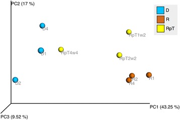
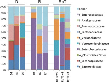
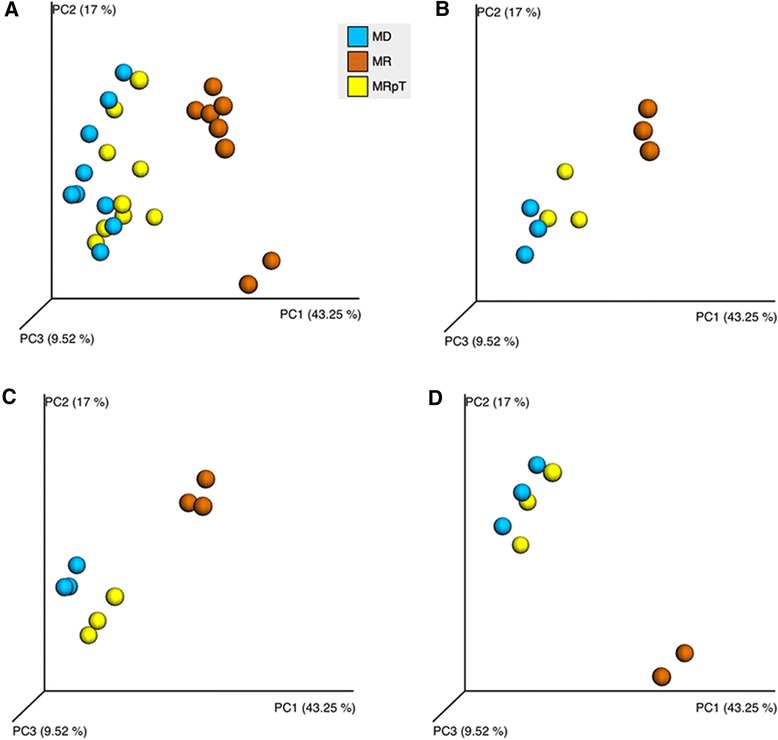
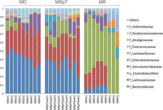
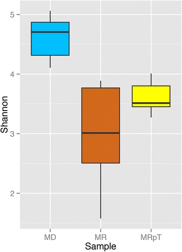
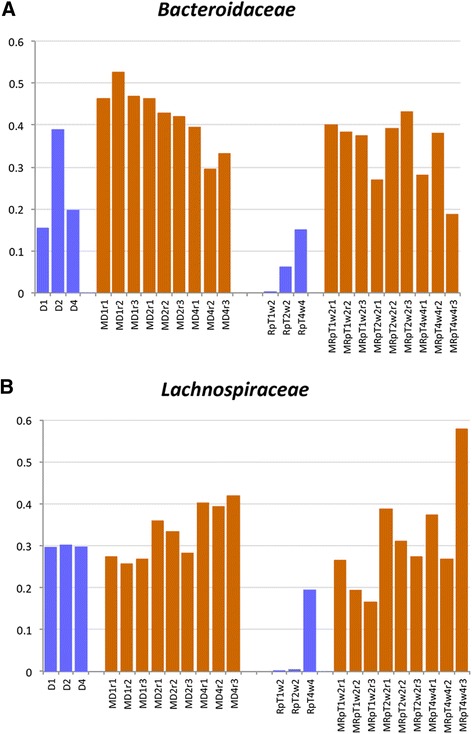
Results: A total of nine samples from three human donors, recipient's pre and post FMT were transplanted into gnotobiotic mice. Microbiome analysis of three donor fecal samples revealed the presence of a high relative abundance of commensal microbes from the family Bacteriodaceae and Lachnospiraceae that were almost absent in the three recipient pre FMT fecal samples (<0.01%). The microbe composition in gnotobiotic mice transplanted with the donor fecal samples was similar to the human samples. The recipient samples contained Enterobacteriaceae, Lactobacillaceae, Enterococcaceae in relative abundance of 43, 11, 8%, respectively. However, gnotobiotic mice transplanted with the recipient fecal samples had an average relative abundance of unclassified Clostridiales of 55%, approximately 7000 times the abundance in the recipient fecal samples prior to transplant. Microbiome analysis of fecal samples from the three patients early (2-4 weeks) after FMT revealed a microbe composition with the relative abundance of both Bacteriodaceae and Lachnospiraceae that was approximately 7% of that of the donor. In contrast, gnotobioitc mice transplanted with the fecal samples obtained from the three at early times post FMT revealed increases in the relative abundance of Bacteriodaceae and Lachnospiraceae microbe compositions to levels similar to the donor fecal samples. Furthermore, the unclassified Clostridiales in the recipient samples post FMT was reduced to an average of 10%.

Conclusion: We have used transplantation into gnotobiotic mice to evaluate the colonization potential of microbiota in FMT patients early after transplant. The commensal microbes present at early times post FMT out competed non-commensal microbes (e.g. such as unclassified Clostridiales) for niche space. The selective advantage of these commensal microbes to occupy niches in the gastrointestinal tract helps to explain the success of FMT to reconstitute the gut microbe community of patients with recurrent C. difficile infections.


Figures

Fig. 1
Fig. 1
Comparison of the microbial diversity of human donors and recipients pre and post FMT. Principal Coordinate analysis is used to generate 3D PCoA plot (using weighted UniFrac distance metrics) for the fecal samples from the donor (D) and recipient (R) and post FMT (RpT)
Fig. 2
Fig. 2
Taxa distribution of human donors and recipients pre and post transplant. A stacked bar plot depicting the taxa distribution at the family level of the fecal samples of individual samples from the donors and recipient pre and post FMT
Fig. 3
Fig. 3
Comparison of the microbiota community in gnotobiotic mice transplanted with fecal samples from donors, recipients pre and post transplant. Gnotobiotic mice were transplanted with the human fecal samples from donor, recipient and RpT samples from FMT 1, 2 and 4. Panel a PCoA plot of the fecal samples from all the gnotobiotic mice. The mice transplanted with donor, recipient, and RpT are named as MD, MR and MRpT, respectively. The orange colored spheres are from mice transplanted with the recipient (1, 2 or 4) samples (MR), the blue spheres are from mice transplanted with donor (1, 2 or 4) (MD) and the yellow spheres are mice transplanted with fecal samples of the FMT (MRpT). Panel b, c and d Individual PCoA plots of donor, recipient and RpT from set 1 (Panel b), 2 (Panel c) and 4 (Pane d) transplanted into gnotobiotic mice. Each panel represents the PCoA plot of individual mice transplanted with the donor and recipient pre and post FMT (RpT)
Fig. 4
Fig. 4
Taxa distribution of the microbial composition of mouse transplanted with donor, recipient pre and post FMT. A stacked bar plot depicting the taxa distribution at the family level of the fecal samples of individual gnotobiotic mice transplanted with samples from the donors and recipient pre and post FMT
Fig. 5
Fig. 5
Shannon diversity for gnotobiotic mice transplanted with donor, recipient pre and post FMT. Alpha diversity (Shannon’s diversity) for gnotobiotic mice transplanted with the donor (blue), recipients pre (orange) and post FMT (yellow) microbiota presented as a box plot
Fig. 6
Fig. 6
Comparison of relative abundance of selected taxa (family level). The human samples are colored in blue and the gntobiotic mice samples are colored orange. Panel a The relative abundance of Bacteroides in the human donors and post FMT and their corresponding transplants in gnotobiotic mice. Panel b The relative abundance of Lachnospiraceae in the human donors and post FMT and their corresponding transplant in gnotobiotic mice

Similar articles

Cited by

References

    1. He M, Miyajima F, Roberts P, Ellison L, Pickard DJ, Martin MJ, et al. Emergence and global spread of epidemic healthcare-associated Clostridium difficile. Nat Genet. 2013;45:109–113. doi: 10.1038/ng.2478. - DOI - PMC - PubMed
    1. Jagai J, Naumova E. Clostridium difficile-associated disease in the elderly, United States. Emerg Infect Dis. 2009;15:343–344. doi: 10.3201/eid1502.080785. - DOI - PMC - PubMed
    1. Kyne L, Hamel MB, Polavaram R, Kelly CP. Health care costs and mortality associated with nosocomial diarrhea due to Clostridium difficile. Clin Infect Dis. 2002;34:346–353. doi: 10.1086/338260. - DOI - PubMed
    1. Seekatz AM, Young VB. Clostridium difficile and the microbiota. J Clin Invest. 2014;124:4182–4189. doi: 10.1172/JCI72336. - DOI - PMC - PubMed
    1. Louie TJ, Miller MA, Mullane KM, Weiss K, Lentnek A, Golan Y, et al. Fidaxomicin versus vancomycin for Clostridium difficile infection. N Engl J Med. 2011;364:422–431. doi: 10.1056/NEJMoa0910812. - DOI - PubMed

Publication types

LinkOut - more resources