. 2016 Mar 15;76(6):1367-80.
doi: 10.1158/0008-5472.CAN-15-1591. Epub 2016 Jan 12.

Neutrophil Extracellular Traps Promote the Development and Progression of Liver Metastases after Surgical Stress

Affiliations

Neutrophil Extracellular Traps Promote the Development and Progression of Liver Metastases after Surgical Stress

Samer Tohme et al. Cancer Res. .

Abstract

Risks of tumor recurrence after surgical resection have been known for decades, but the mechanisms underlying treatment failures remain poorly understood. Neutrophils, first-line responders after surgical stress, may play an important role in linking inflammation to cancer progression. In response to stress, neutrophils can expel their protein-studded chromatin to form local snares known as neutrophil extracellular traps (NET). In this study, we asked whether, as a result of its ability to ensnare moving cells, NET formation might promote metastasis after surgical stress. Consistent with this hypothesis, in a cohort of patients undergoing attempted curative liver resection for metastatic colorectal cancer, we observed that increased postoperative NET formation was associated with a >4-fold reduction in disease-free survival. In like manner, in a murine model of surgical stress employing liver ischemia-reperfusion, we observed an increase in NET formation that correlated with an accelerated development and progression of metastatic disease. These effects were abrogated by inhibiting NET formation in mice through either local treatment with DNAse or inhibition of the enzyme peptidylarginine deaminase, which is essential for NET formation. In growing metastatic tumors, we found that intratumoral hypoxia accentuated NET formation. Mechanistic investigations in vitro indicated that mouse neutrophil-derived NET triggered HMGB1 release and activated TLR9-dependent pathways in cancer cells to promote their adhesion, proliferation, migration, and invasion. Taken together, our findings implicate NET in the development of liver metastases after surgical stress, suggesting that their elimination may reduce risks of tumor relapse.


Conflict of interest statement

Conflict of interest: K. Mowen reports employment with, research support from, and service on the board of Padlock Therapeutics and a commercial research grant from Janssen. The remaining authors have declared that no conflict of interest exists.

Figures

Figure 1
Figure 1
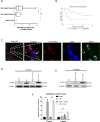
Surgical stress induced by liver I/R results in widespread deposition of NETs. (A) Detection of serum MPO-DNA levels in mCRC patients undergoing major (n=35) or minor liver surgery (n=15) or in healthy controls (n=20). Box plots show higher MPO-DNA levels in mCRC patients undergoing major resection. (B) Kaplan-Meier disease-free survival curves of mCRC patients who underwent major liver resection based on serum MPO-DNA levels. (C) Representative immunofluorescence images by confocal microscopy of mice liver sections showing NETs 60 min after ischemia/tumor injection and 6 h reperfusion (magnification 40×, n=6) with staining for Ly6G(red), nuclei(blue), histone H2AX(green), F-actin(gray). Arrow: released histone and DNA from neutrophils. (D and E) Cit-H3 protein levels were determined by Western blot in sham, sham+tumor injection, I/R, and I/R+tumor injection mice groups after liver I/R at (D) 6 hours and (E) 3 weeks. Hepatic protein lysates from ischemic lobes at 6 hours or tumors at 3 weeks were obtained. The blots shown are representatives of three experiments with similar results. (F) NETs acutely form in liver tissue 6 hours after tumor injection and liver I/R, and in tumors 3 weeks later, as assessed by serum levels of MPO-DNA. Results are expressed as the relative folds increase of MPO-DNA levels compared with sham; mean±SEM (n=6/group). *P<0.05, ***P<0.001.
Figure 2
Figure 2
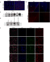
Growing Solid Tumors generate a hypoxic environment that potentiated NET formation. (A) Three weeks after splenic injection of MC38 cells, the liver tumors exhibit significant central hypoxia as evident by increased HIF-1α staining by immunofluorescent imaging compared to background liver. HIF1-α (red), nuclei (Blue) (B) Using confocal microscopy, there is a significant increase in tumor infiltrating neutrophils three weeks after mice were subjected to I/R compared to mice that only received tumor injection surgery (mean 258 Ly6G+ cells/106μm2 versus 20 Ly6G+ cells/106μm2, p<0.001). Ly6G (red), nuclei (blue). Scale Bars A and B: 100μm. (C) Cit-H3 levels, marker of NETs, was increased in the hypoxic liver tumors compared to background liver in paired mouse tissues three weeks after establishment of the metastatic and liver I/R model. (D and E) In vitro, there was a significant increase in NET formation after neutrophils isolated from mice were co-cultured with media from MC38 cells subjected to hypoxia or thermal necrosis as evident by (D) Western blot for cit-H3 and (E) confocal microscopy at 40× magnification. Cit-H3 (green), Nuclei (blue), F-actin (red). Arrows indication neutrophils forming NETs. (N) Normal/T (Tumor)
Figure 3
Figure 3
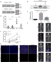
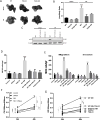
Surgical stress promotes the development of gross metastases, which is attenuated by administration of DNAse, a NET inhibitor. (A) A schematic representation of the experimental design is depicted. Mice were subjected to liver I/R in order to induce surgical stress. Intrasplenic injection of MC38 colorectal cancer cell lines was performed at the same time. Daily DNAse intraperitoneally was started at the time of procedure. (B) Cit-H3 levels were significantly decreased after administration of DNAse after 1 hour of ischemia and 6 hours of reperfusion. (C) Treatment with DNAse after liver I/R resulted in a significant decrease in the levels of serum MPO-DNA at 6 hours. (D) At 14 days, I/R resulted in a significant increase in gross surface metastatic nodules compared with the sham groups weeks (mean 11 nodules in I/R versus 2 nodules in sham; p<0.0001). Treatment with DNAse resulted in a significant decrease in the number of gross metastases. (E and F) The use of luciferase-labeled MC38 cells allowed weekly in vivo tracking of tumor growth with bioluminescence imaging. DNase does not affect tumor growth in the unstressed tumors, the sham group. In the I/R group, we again demonstrate the effect of surgical stress on the acceleration of tumor growth and this is significantly inhibited at day 7 and 14 by the daily administration of DNAse. Data represent mean±SEM; n=6 mice/group. The above data are each representative of three experiments with similar results. (G) Daily treatment with DNAse after I/R significantly attenuated tumor cell proliferation weeks (mean 22 Ki67+ cells/106μm2 I/R and DNase group versus 96 Ki67+ cells/106μm2 in I/R group, p<0.001) and tumor associated tumorigenesis (mean CD31+ area 6×103μm2 versus 18×103μm2/106μm2, p<0.001) in liver metastatic tumors at three. Blue (nuclei), Ki67 (green), Cd31 (red); Scale bar=500μm. NS: not significant, **P<0.01, ***P<0.001.
Figure 4
Figure 4
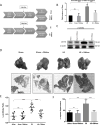
Surgical stress promotes the growth of established liver micrometastases, which is attenuated by administration of DNAse. (A) Schematic representation of the experimental design is depicted. Intrasplenic injection of MC38 cells was performed and metastatic tumor was allowed to grow for 5 days before the mice were subjected to liver I/R. Daily DNAse treatment was started at the time of liver I/R. At 3 weeks, the mice were sacrificed and tumor growth was assessed. (B and C) There was a significant increase in NET formation in the tumors after I/R as evident by increase in (B) tumor cit-H3 levels and (C) serum levels of MPO-DNA. Daily DNAse treatment resulted in a significant inhibition NET formation. (D) Representative images of hepatic nodules and H&E staining of liver section (N=normal, T=tumor) after necropsy in mice subjected to sham or I/R with or without daily DNAse treatment. (E and F) Liver I/R resulted in a significant increase in tumor burden at three weeks as seen by (E) liver-to-body ratio (45% decrease with DNAse treatment after I/R, p<0.001) and (F) percentage hepatic replacement by metastatic tumor (mean percentage replacement 78% in I/R vs 44% in I/R plus DNAse, p=0.02). Treatment with DNAse after I/R resulted in a significant decrease in growth of already established micrometastases. Data represent mean±SEM; n=6 mice/group. The above data are each representative of three experiments with similar results. NS: not significant, ***P<0.001.
Figure 5
Figure 5
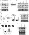
DNAse and PAD4 targeting inhibit the pro-tumorigenic effects of NETs. MC38 cancer cells were injected in the spleen of C57BL/6 wild type (WT) mice or PAD4 KO mice with or without liver I/R. In addition, a subgroup of WT mice was injected intraperitoneally three times/week with PAD4 inhibitor YW04-03. Mice deficient in PAD4 or WT mice treated with YW4-03 had a significant decrease in tumor burden at three weeks as demonstrated by the representative images (A) and the liver-to-body ratio (B) compared to WT mice after I/R. (C) There was a significant decrease in NET formation in the tumors after I/R in PAD4 deficient mice and in mice treated with YW4-03 as evident by decrease in tumor cit-H3 levels. (D) In vitro, CFSE-labelled MC38 cells demonstrated increased adhesion to neutrophil monolayers stimulated with PMA (100nM) compared with unstimulated neutrophils or neutrophils from PAD4 deficient mice (mean 19.1 MC38 cells/hpf versus 5.7 MC38 cells/hpf versus 6.2 MC38 cells/hpf; p<0.0001). Addition of DNAse or YW4-03 results in adhesion levels comparable to control (mean 8.1 and 7.2 MC38 cells/hpf, respectively; p<0.0001 compared to PMA treatment). (E) MC38 cell migration through 8-μm PET membranes and invasion through Matrigel was significantly increased in the presence of media from PMA-stimulated neutrophils (NM) compared with MC38 alone. This was reversed back to control values with the addition of DNAse or YW3-04 to the stimulated media or when using stimulated media from PAD4 deficient mice. (F and G) MTT assay show increased tumor proliferation at 48 hours after culturing MC38 cells with media from PMA-stimulated neutrophils (NM) but not from PMA-stimulated neutrophils from PAD4 deficient mice. Addition of DNAse or YW4-03 result in proliferation levels similar to control. Data are presented as mean±SEM from n=3 separate experiments. NS: not significant, ***P<0.001.
Figure 6
Figure 6
NETs exert their pro-tumorigenic effects through activation of TLR-9 pathways. (A) At three weeks, MAP kinase pathways were increasingly activated in the tumors obtained from mice that underwent I/R and tumor injection (B) In vitro, TLR9 expression is significantly increased after MC38 cancer cells were cultured in media derived from PMA-stimulated neutrophils (NM). (C) The addition of NM significantly increase the activation of the TLR9 associated MAP kinases compared to control; this was similar to the addition of a TLR9 agonist (ODN 1668). The addition of DNAse to NM resulted in phosphorylation levels similar to control. Furthermore, the addition of NM had no effect in TLR9-knockdown MC38 cells. (D and E) Similarly, the addition of NM did not affect tumor cell (D) proliferation, (E) migration or invasion in TLR9 knocked down MC38 cells when compared to effect of NM in wild type MC38 cells. (F) The addition of NM failed to activate TLR9 pathways when neutrophils were pretreated with YW4-03 or when using PAD4 deficient neutrophils. (G and H) In vivo, mice injected with MC38 cells deficient in TLR9 had a significant decrease in tumor growth in response to I/R at three weeks as demonstrated by the representative images (H) and the liver-to-body ratio (I) compared to mice injected with WT MC38 cells. (I) As expected, there was not an increase in MAP kinase pathway activation after I/R in TLR9 knocked down MC38 cells. *P<0.0001 versus control; **P<0.001, ***P<0.01 versus NM. Data in C, D, E are presented as mean ± SEM from n=3 separate experiments; in vivo experiments (n=6/group, tumor protein lysate were obtained, each lane represents a separate animal). NS: not significant, **P<0.01, ***P<0.001.
Figure 7
Figure 7
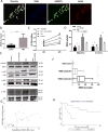
HMGB1 released from NETs promote tumor progression and TLR9 activation. (A) After PMA stimulation for 4 hours, confocal microscopy images of NETs reveal that HMGB1 is released and co-localizes with extruded DNA. Ly6G (red), nuclei (blue), HMGB1 (green). (B) Using ELISA, HMGB1 levels are increased in the supernatant from PMA-stimulated neutrophils compared to unstimulated neutrophils. (C and D) The addition of neutralizing HMGB1 antibodies substantially reversed the effect of NM of MC38 cell (C) proliferation, (D) migration and invasion. (E) MC38 cells were cultured in media collected from PMA-stimulated neutrophils (NM). The addition of neutralizing HMGB1 antibodies to the NM reversed the activation of the MAP kinases back to control level. Data are presented as mean ± SEM from n=3 separate experiments. *P<0.05, ***P<0.001. (F) Detection of preoperative and postoperative serum HMGB1 levels in mCRC patients undergoing minor or major surgery (n=50) or in healthy controls (n=20). Box plots show higher HMGB1 levels in mCRC patients compared to healthy controls and higher HMGB1 levels postoperatively compared to preoperatively. (G) Postoperative HMGB1 levels correlated with increases in serum MPO-DNA levels. (H) Kaplan-Meier disease-free survival curves of mCRC patients who underwent major liver resection based on serum HMGB1 levels.

References

    1. Siegel RL, Miller KD, Jemal A. Cancer statistics, 2015. CA: a cancer journal for clinicians. 2015 Jan-Feb;65(1):5–29. - PubMed
    1. Kopetz S, Chang GJ, Overman MJ, Eng C, Sargent DJ, Larson DW, et al. Improved survival in metastatic colorectal cancer is associated with adoption of hepatic resection and improved chemotherapy. Journal of clinical oncology : official journal of the American Society of Clinical Oncology. 2009 Aug 1;27(22):3677–83. - PMC - PubMed
    1. Wolpin BM, Mayer RJ. Systemic treatment of colorectal cancer. Gastroenterology. 2008 May;134(5):1296–310. - PMC - PubMed
    1. Murthy SM, Goldschmidt RA, Rao LN, Ammirati M, Buchmann T, Scanlon EF. The influence of surgical trauma on experimental metastasis. Cancer. 1989 Nov 15;64(10):2035–44. - PubMed
    1. van der Bij GJ, Oosterling SJ, Beelen RH, Meijer S, Coffey JC, van Egmond M. The perioperative period is an underutilized window of therapeutic opportunity in patients with colorectal cancer. Annals of surgery. 2009 May;249(5):727–34. - PubMed

Publication types

MeSH terms