. 2016 May;36(5):917-27.
doi: 10.1177/0271678X15625578. Epub 2016 Jan 12.

Ischemic penumbra as a trigger for intracranial pressure rise - A potential cause for collateral failure and infarct progression?

Affiliations

Ischemic penumbra as a trigger for intracranial pressure rise - A potential cause for collateral failure and infarct progression?

Daniel J Beard et al. J Cereb Blood Flow Metab. 2016 May.

Abstract

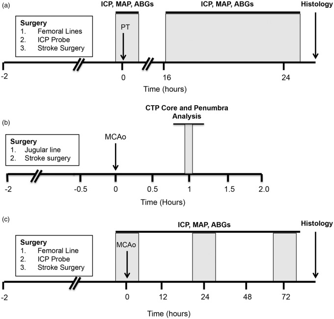
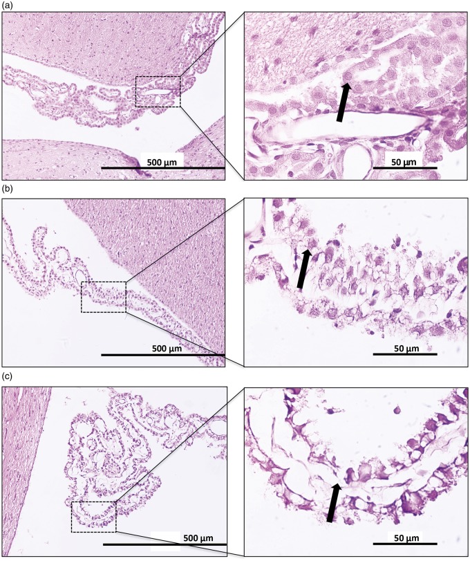
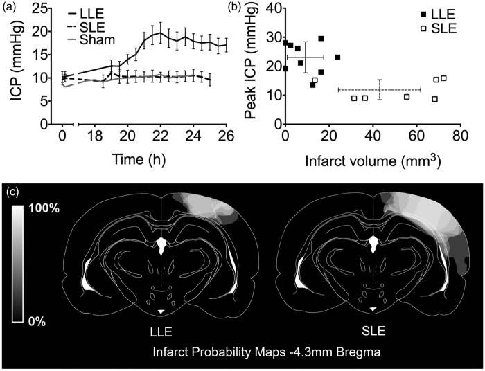
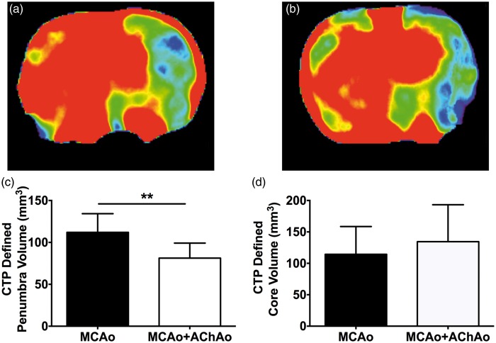
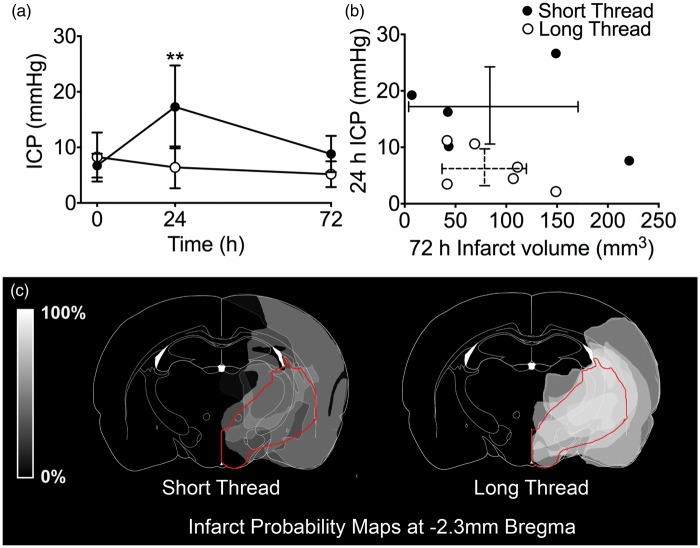
We have recently shown that intracranial pressure (ICP) increases dramatically 24 h after minor intraluminal thread occlusion with reperfusion, independent of edema. Some of the largest ICP rises were observed in rats with the smallest final infarcts. A possible alternate mechanism for this ICP rise is an increase of cerebrospinal fluid (CSF) volume secondary to choroid plexus damage (a known complication of the intraluminal stroke model used). Alternatively, submaximal injury may be needed to induce ICP elevation. Therefore, we aimed to determine (a) if choroid plexus damage contributes to the ICP elevation, (b) if varying the patency of an important internal collateral supply to the middle cerebral artery (MCA), the anterior choroidal artery (AChA), produces different volumes of ischemic penumbra and (c) if presence of ischemic penumbra (submaximal injury) is associated with ICP elevation. We found (a) no association between choroid plexus damage and ICP elevation, (b) animals with a good internal collateral supply through the AChA during MCAo had significantly larger penumbra volumes and (c) ICP elevation at ≈24 h post-stroke only occurred in rats with submaximal injury, shown in two different stroke models. We conclude that active cellular processes within the ischemic penumbra may be required for edema-independent ICP elevation.

Keywords: Collaterals; intracranial pressure; middle cerebral artery occlusion; penumbra; photothrombosis.


Figures

Figure 1.
Figure 1.
Experimental timelines. (a) Study I. ICP was monitored at baseline and between 18 and 26 h after PT stroke. (b) Study II. CTP imaging was carried out to measure whole brain perfusion changes during MCAo. For the purposes of this study, scans taken 1 h post-MCAo were used to create core and penumbra maps using previously established CTP-defined CBF thresholds for this model. (c) Study III. ICP was monitored at baseline, 24 and 72 h after permanent MCAo. ABG = arterial blood gas; CTP: computed tomography perfusion; ICP: intracranial pressure; MAP: mean arterial pressure; MCAo: middle cerebral artery occlusion; PT: photothrombotic stroke.
Figure 2.
Figure 2.
Choroid plexus damage scoring. Hematoxylin and eosin staining of lateral ventricle choroid plexus at 20× (left panels) and 100× (right panels). (a) Normal choroid plexus histology (given a score of 0) showing a continuous epithelial lining of the choroidal villus with healthy epithelial cells (arrow). (b) Moderate choroid plexus damage (given a score of 1) showing an intact epithelial lining with shrunken and vacuolated epithelial cells (arrow). (c) Severe choroid plexus damage (given a score of 2) showing areas of epithelial desquamation (arrow).
Figure 3.
Figure 3.
Study I – ICP, CPP and infarct volume following photothrombotic stroke. (a) ICP 0–0.5 h and 18–26 h post-photothrombotic stroke in Wistar rats using either LLE (black line; n = 9) or SLE (broken black line; n = 7) and sham animals (grey lines; n = 2), mean ± SEM. Each data point represents ICP averaged over 10 min, at 30-min time intervals. (b) Peak ICP versus 24-h infarct volume for LLE (filled squares, unbroken error bars) or SLE (open squares, broken error bars). (c) Infarct probability map at −4.3 mm Bregma, where the maximal extent of the photothrombotic lesions were located. Lighter regions represent areas more commonly infarcted. CPP: cerebral perfusion pressure; ICP: intracranial pressure; LLE: low light exposure; SEM: standard error mean; SLE: standard light exposure.
Figure 4.
Figure 4.
Study II – CTP core and penumbra volume analysis in animals with or without concomitant AChAo during intraluminal MCAo. Representative 1-h CBF maps for animals with MCAo (a) or MCAo + AChA (b). Penumbra volume (c), core volume (d) at 1 h post-MCAo in animals with MCAo only (black bars) and animals with MCAo + AChAo (white bars). **p < 0.01. AChA: anterior choroidal artery; AChAo: anterior choroidal artery occlusion; CTP: computer tomography perfusion; MCAo: middle cerebral artery occlusion.
Figure 5.
Figure 5.
Study III – ICP, infarct volume and infarct probability following pMCAo in Wistar rats. (a) ICP at baseline, 24 h and 72 h post-pMCAo. Strokes were induced with intraluminal threads with silicon tip length designed either to maintain AChA patency (1.5 mm tips, short thread group, closed circles; n = 6) or to occlude the AChA (4 mm tips, long thread group, open circles; n = 6). Main effect of thread length on ICP, F (1,3) = 6.33, p = 0.023; ANCOVA. **p < 0.01 between groups; post-hoc Bonferroni t-tests (b) Relationship between 72-h infarct volume and 24-h ICP in the short thread group (closed circles, unbroken error bars) and the long thread group (open circles, broken error bars). (c) Infarct probability map at −2.3 mm Bregma. Lighter regions represent areas more commonly infarcted. The region supplied by the AChA is outlined in red. AChA: anterior choroidal artery; ANCOVA: analysis of covariance: ICP: intracranial pressure; pMCAo: permanent middle cerebral artery occlusion.

References

    1. Murtha LA, McLeod DD, McCann SK, et al. Short-duration hypothermia after ischemic stroke prevents delayed intracranial pressure rise. Int J Stroke 2014; 9: 553–559. - PubMed
    1. Murtha LA, McLeod DD, Pepperall D, et al. Intracranial pressure elevation after ischemic stroke in rats: cerebral edema is not the only cause, and short-duration mild hypothermia is a highly effective preventive therapy. J Cereb Blood Flow Metab 2015; 35: 592–600. - PMC - PubMed
    1. Burrows G. On disorders of the cerebral circiulation and on the connection between affections of the brain and diseases of the heart, London: Longman, 1846. - PMC - PubMed
    1. Ennis SR, Keep RF. The effects of cerebral ischemia on the rat choroid plexus. J Cereb Blood Flow Metab 2006; 26: 675–683. - PubMed
    1. Coutts SB, Modi J, Patel SK, et al. CT/CT angiography and MRI findings predict recurrent stroke after transient ischemic attack and minor stroke: results of the prospective CATCH study. Stroke 2012; 43: 1013–1017. - PubMed

Publication types

LinkOut - more resources