

Application of direct oral microscopy in evaluating mucosal margins around invasive oral squamous cell carcinoma
- PMID: 26759543
- PMCID: PMC4692809
- DOI: 10.5114/pdia.2014.40792
Application of direct oral microscopy in evaluating mucosal margins around invasive oral squamous cell carcinoma
Abstract
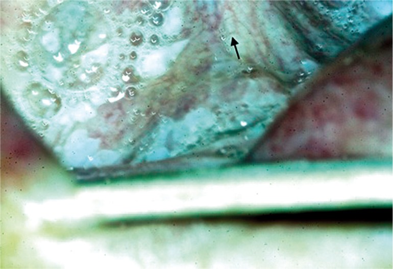
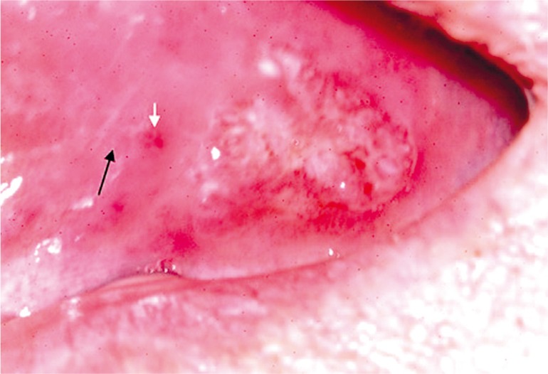
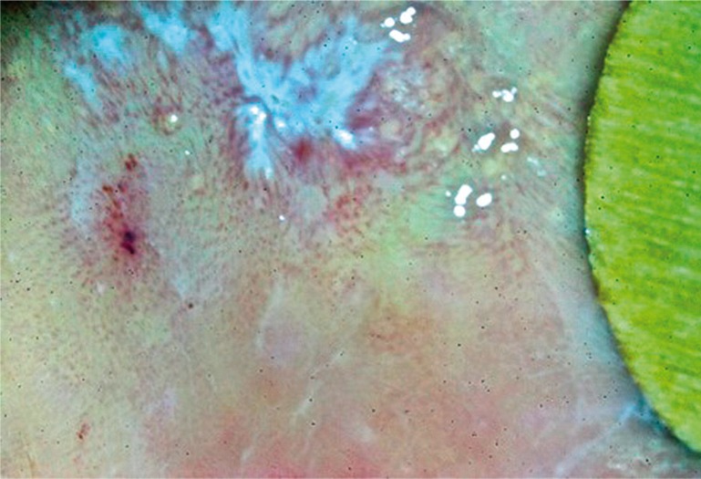
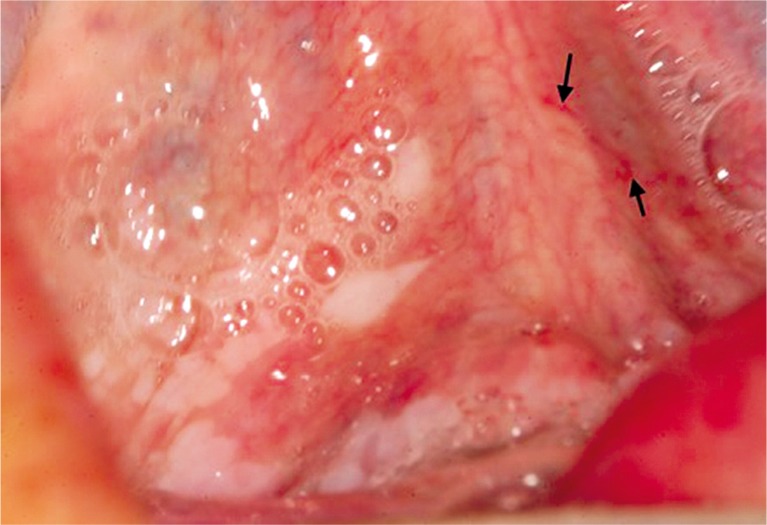
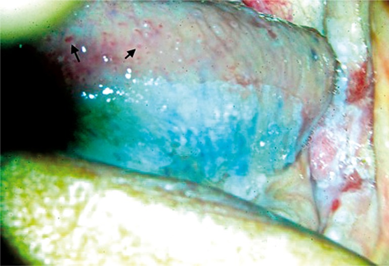
Introduction: Direct oral microscopy constitutes a novel, non-invasive diagnostic technique, which aids clinical examination of the oral cavity. The oral mucosa is examined at multiple magnifications and features such as sub-epithelial mucosal vessels, surface patterns, colour tone, transparency and the exact demarcation of mucosal lesions are estimated. The incidence of oral squamous cell carcinoma (OSCC) oscillates between 1.9% and 3.5%, which makes it the eighth most common carcinoma occurring around the world and in Poland. The 5-year survival rates oscillate between 20% and 30%.
Aim: The aim of the study was to evaluate clinically unchanged mucosal margins around OSCC by direct oral microscopy. The authors approached the question whether the borders of mucosal margins around OSCC established via direct oral microscopy differ from those established based on clinical examination.
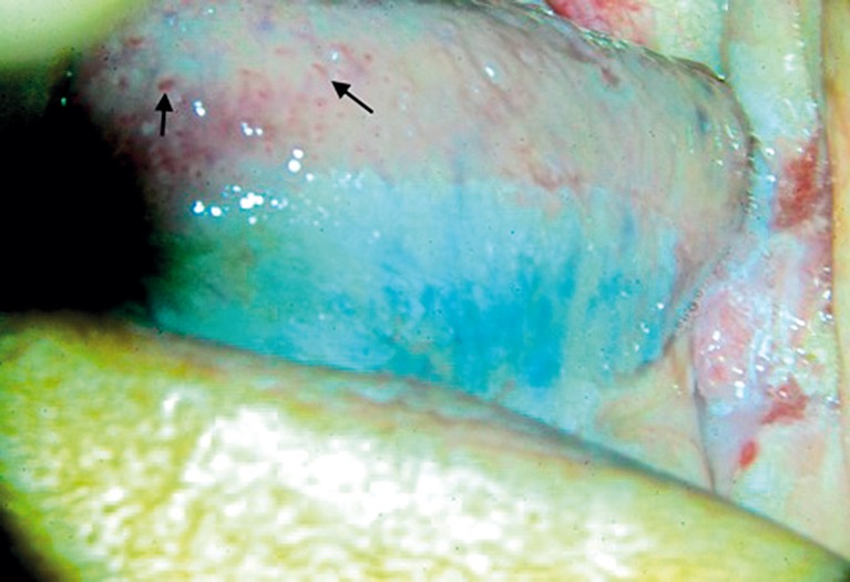
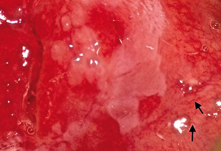
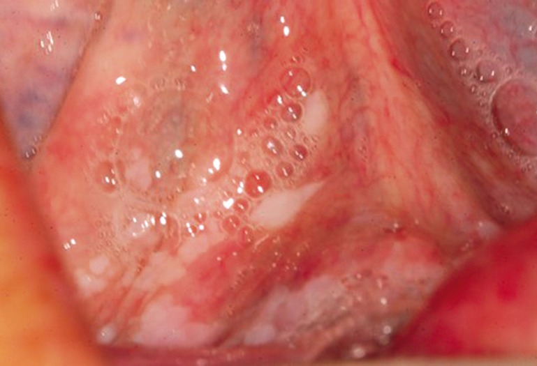
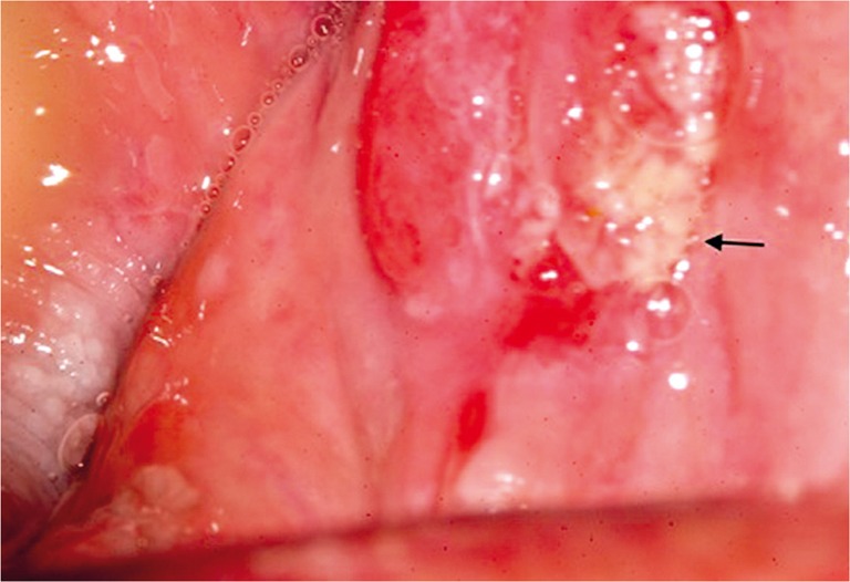
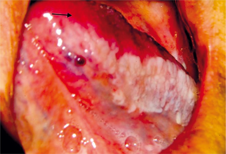
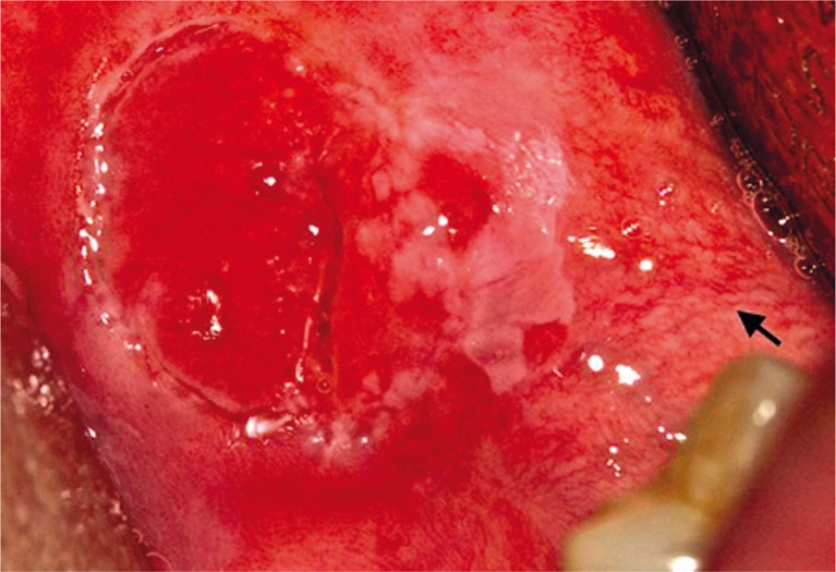
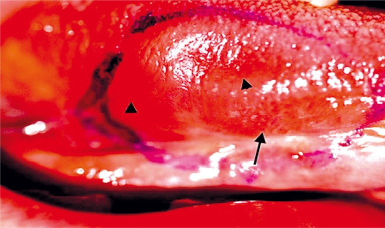
Material and methods: Fifteen patients diagnosed with OSCC were enrolled. Patients were first clinically examined to evaluate the extent of the tumour and to plan resection margins. Eventually, direct oral microscopy was performed to establish the width of the subclinically unchanged mucosal margins based on a standard picture of healthy oral mucosae, followed by comparison with those established by clinical evaluation.
Results: Histopathologic results of biopsies from areas indicated by direct oral microscopy revealed dysplasia in 86.7% of patients, whereas biopsies from areas indicated by clinical examination revealed dysplasia only in 40% of individuals, resulting in the need for widening of mucosal margins.
Conclusions: Direct oral microscopy enables detection of dysplasia within clinically unaltered mucosal margins around OSCC, which results in more precise establishing of resection boundaries, contributing to improvement of resection totality.
Keywords: direct microscopy; oral cavity; squamous cell carcinoma.
Figures
Similar articles
-
A standard picture of healthy oral mucosae by direct oral microscopy.Postepy Dermatol Alergol. 2013 Jun;30(3):159-64. doi: 10.5114/pdia.2013.35617. Epub 2013 Jun 20. Postepy Dermatol Alergol. 2013. PMID: 24278068 Free PMC article.
-
Clinical diagnosis of oral erosive lichen planus by direct oral microscopy.Postepy Dermatol Alergol. 2014 Aug;31(4):222-8. doi: 10.5114/pdia.2014.40926. Epub 2014 Sep 8. Postepy Dermatol Alergol. 2014. PMID: 25254007 Free PMC article.
-
Identification of salivary metabolites for oral squamous cell carcinoma and oral epithelial dysplasia screening from persistent suspicious oral mucosal lesions.Clin Oral Investig. 2019 Sep;23(9):3557-3563. doi: 10.1007/s00784-018-2777-3. Epub 2018 Dec 11. Clin Oral Investig. 2019. PMID: 30539290
-
AGA Institute Clinical Practice Update: Endoscopic Submucosal Dissection in the United States.Clin Gastroenterol Hepatol. 2019 Jan;17(1):16-25.e1. doi: 10.1016/j.cgh.2018.07.041. Epub 2018 Aug 2. Clin Gastroenterol Hepatol. 2019. PMID: 30077787 Review.
-
Molecular diagnostics in oral cancer and oral potentially malignant disorders-A clinician's guide.J Oral Pathol Med. 2020 Jan;49(1):1-8. doi: 10.1111/jop.12920. Epub 2019 Aug 3. J Oral Pathol Med. 2020. PMID: 31309636 Review.
References
-
- Gynther GW, Rozell B, Heimdahl A. Direct oral microscopy and its value in diagnosing mucosal lesions. A pilot study. Oral Surg Oral Med Oral Pathol Oral Radiol Endod. 2000;90:164–70. - PubMed
-
- Xiumei W, Wenjing S, Jing B, et al. Growth inhibition induced by transforming growth factor beta1 in human oral squamous cell carcinoma. Mol Biol Rep. 2009;36:861–9. - PubMed
-
- Strycharz M, Polz-Dacewicz M, Gołąbek W, et al. The epidemiologic analysis of 254 oral cancer cases from the Lublin region. Lublin, Polonia: Annales Universitatis Mariae Curie-Skłodowska; 2006. pp. 655–9. N 2, 114, Sectio D.
-
- Rigual NR, Wiseman SM. Neck dissection: current concepts and future directions. Surg Oncol Clin N Am. 2004;13:151–66. - PubMed
LinkOut - more resources
Full Text Sources
Other Literature Sources