

C1 anterior arch preservation in transnasal odontoidectomy using three-dimensional endoscope: A case report
- PMID: 26759737
- PMCID: PMC4697203
- DOI: 10.4103/2152-7806.172696
C1 anterior arch preservation in transnasal odontoidectomy using three-dimensional endoscope: A case report
Abstract
Background: The transoral ventral corridor is the most common approach used to reach the craniovertebral junction (CVJ). Over the last decade, many case reports have demonstrated the transnasal corridor to the odontoid peg represents a practicable route to remove the tip of the odontoid process. The biomechanical consequences of the traditional odontoidectomy led to the necessity of a cervical spine stabilization. Preserving the inferior portion of the C1 anterior arch should prevent instability.
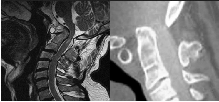
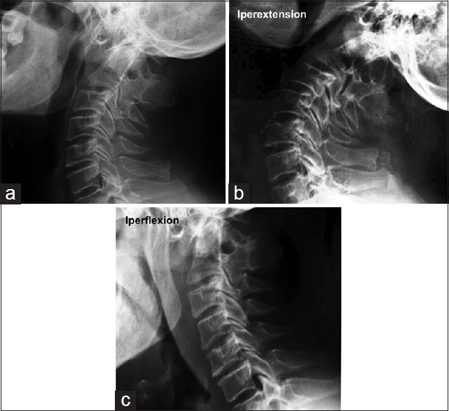
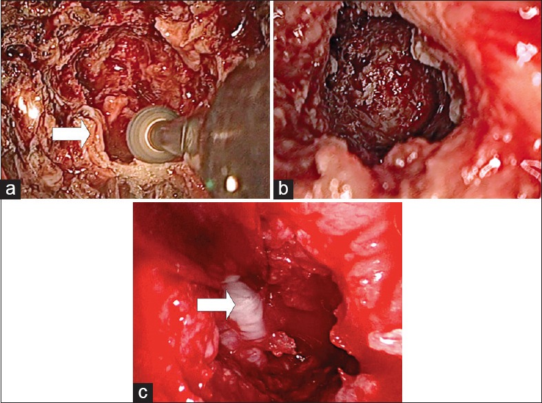
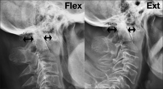
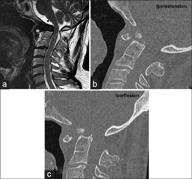
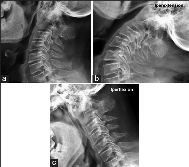
Case description: This is the first report in which the technique to remove the tip of the odontoid while preserving the C1 anterior arch is described by means of a three-dimensional (3D) endoscope. A 53-year-old man underwent a transnasal 3D endoscopic approach because of a complex CVJ malformation. The upper-medial portion of the C1 anterior arch was removed preserving its continuity, and the odontoidectomy was performed. After surgery, a dynamic X-ray scan showed no difference in CVJ motility in comparison with the preoperative one.
Conclusions: The stereoscopic perception augmented the precision of the surgical gesture in the deep field. The importance of a 3D view relates to the depth of field, which a two-dimensional endoscopy cannot provide. This affects the preservation of the C1 anterior arch because of the presence of critical structures that are exposed to potential damage if not displayed.
Keywords: C1 anterior arch preservation; craniovertebral junction stability; three-dimensional endoscope; transnasal odontoidectomy.
Figures
References
-
- Agrawal A, Reyes PM. A novel technique of odontoidoplasty and C1 arch reconstruction: Anatomical and biomechanical basis. Neurosurgery. 2011;68(1 Suppl Operative):103–13. - PubMed
-
- Alfieri A, Jho HD, Tschabitscher M. Endoscopic endonasal approach to the ventral cranio-cervical junction: Anatomical study. Acta Neurochir (Wien) 2002;144:219–25. - PubMed
-
- Blazier CJ, Hadley MN, Spetzler RF. The transoral surgical approach to craniovertebral pathology. J Neurosci Nurs. 1986;18:57–62. - PubMed
-
- Cappabianca P, Cavallo LM, Esposito I, Barakat M, Esposito F. Bone removal with a new ultrasonic bone curette during endoscopic endonasal approach to the sellar-suprasellar area: Technical note. Neurosurgery. 2010;66:E118. - PubMed
Publication types
LinkOut - more resources
Full Text Sources
Other Literature Sources