. 2015 Aug 24;4(4):e391-6.
doi: 10.1016/j.eats.2015.05.005. eCollection 2015 Aug.

Basic Hip Arthroscopy: Supine Patient Positioning and Dynamic Fluoroscopic Evaluation

Affiliations

Basic Hip Arthroscopy: Supine Patient Positioning and Dynamic Fluoroscopic Evaluation

Sandeep Mannava et al. Arthrosc Tech. .

Abstract

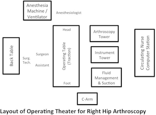
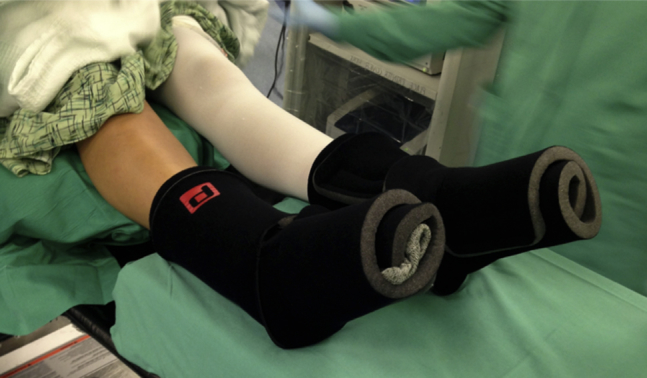
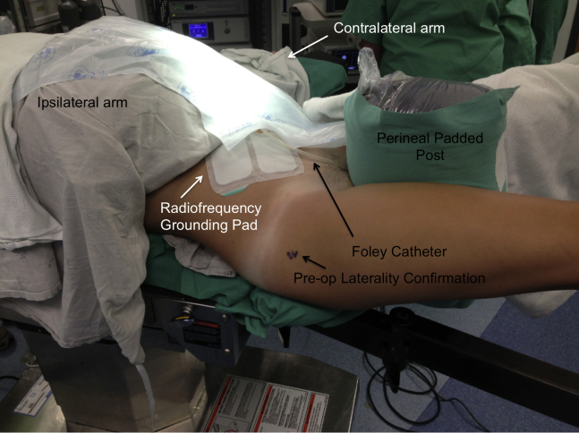
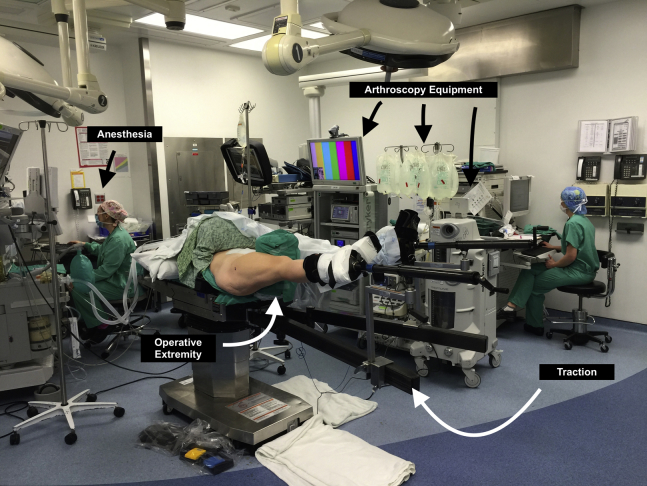
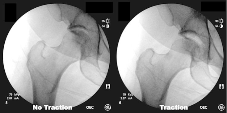
Hip arthroscopy serves as both a diagnostic and therapeutic tool for the management of various conditions that afflict the hip. This article reviews the basics of hip arthroscopy by demonstrating supine patient positioning, fluoroscopic evaluation of the hip under anesthesia, and sterile preparation and draping. Careful attention to detail during the operating theater setup ensures adequate access to the various compartments of the hip to facilitate the diagnosis of disease and treatment with minimally invasive arthroscopy. Furthermore, having a routine method for patient positioning and operative setup improves patient safety, as well as operative efficiency, as the operative team becomes familiar with the surgeon's standard approach to hip arthroscopy cases.


Figures

Fig 1
Fig 1
Operative theater setup for right-sided hip arthroscopy. The locations of various instruments and equipment in the operating theater are shown. The surgeon, assistants, and instrumentation are located on the operative (right) side of the patient. The arthroscopy tower and associated equipment are located on the contralateral (left) side of the patient. (Surg Tech, surgical technologist.)
Fig 2
Fig 2
Bilateral feet after placement of commercially available foam-padded boots for right hip arthroscopy. The contralateral (left) leg is depicted with a compression hose to prevent deep vein thrombosis. The ipsilateral, operative (right) leg does not have a compression hose. Care is taken to avoid wrinkles while applying the foam-padded boots to prevent decubitus ulcers.
Fig 3
Fig 3
Positioning of operative right hip after placement into traction boots and positioning of perineal padded post for right hip arthroscopy. The arms have been positioned so that the ipsilateral (right) arm is placed over the patient and is well padded, and a papoose wrapping has been applied. The contralateral (left) arm is placed on a standard arm board. The perfusion of both upper extremities is confirmed after positioning. The positioning of the Foley catheter and the radiofrequency grounding pad is also shown.
Fig 4
Fig 4
Positioning of patient in traction before sterile preparation and draping for right hip arthroscopy. The operative hip is positioned at 15° of flexion and 0° of adduction before the application of traction. Internal rotation of the foot to 10° to 15° positions the femoral neck more parallel to the operative horizon.
Fig 5
Fig 5
Fluoroscopic views of the right hip before and after application of traction, confirming adequate distraction of the hip joint through the appearance of the crescent sign. If the surgeon is unable to attain the vacuum crescent sign, the joint can be vented with a spinal needle to break the suction seal or the surgeon can continue to add gentle traction until the crescent sign appears.

References

    1. Colvin A.C., Harrast J., Harner C. Trends in hip arthroscopy. J Bone Joint Surg Am. 2012;94:e23. - PubMed
    1. Larson C.M., Stone R.M. Current concepts and trends for operative treatment of FAI: Hip arthroscopy. Curr Rev Musculoskelet Med. 2013;6:242–249. - PMC - PubMed
    1. Montgomery S.R., Ngo S.S., Hobson T. Trends and demographics in hip arthroscopy in the United States. Arthroscopy. 2013;29:661–665. - PubMed
    1. Bozic K.J., Chan V., Valone F.H., III, Feeley B.T., Vail T.P. Trends in hip arthroscopy utilization in the United States. J Arthroplasty. 2013;28(suppl):140–143. - PubMed
    1. Burman M.S. Arthroscopy or the direct visualization of joints: An experimental cadaver study. 1931. Clin Orthop Relat Res. 2001;(390):5–9. - PubMed

LinkOut - more resources