

Topography of acute stroke in a sample of 439 right brain damaged patients
- PMID: 26759787
- PMCID: PMC4683427
- DOI: 10.1016/j.nicl.2015.11.012
Topography of acute stroke in a sample of 439 right brain damaged patients
Abstract
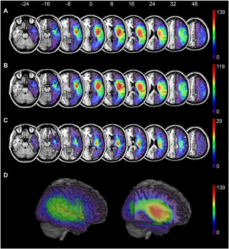
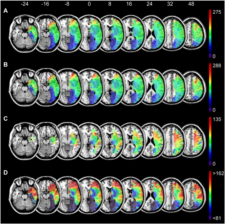
Knowledge of the typical lesion topography and volumetry is important for clinical stroke diagnosis as well as for anatomo-behavioral lesion mapping analyses. Here we used modern lesion analysis techniques to examine the naturally occurring lesion patterns caused by ischemic and by hemorrhagic infarcts in a large, representative acute stroke patient sample. Acute MR and CT imaging of 439 consecutively admitted right-hemispheric stroke patients from a well-defined catchment area suffering from ischemia (n = 367) or hemorrhage (n = 72) were normalized and mapped in reference to stereotaxic anatomical atlases. For ischemic infarcts, highest frequencies of stroke were observed in the insula, putamen, operculum and superior temporal cortex, as well as the inferior and superior occipito-frontal fascicles, superior longitudinal fascicle, uncinate fascicle, and the acoustic radiation. The maximum overlay of hemorrhages was located more posteriorly and more medially, involving posterior areas of the insula, Heschl's gyrus, and putamen. Lesion size was largest in frontal and anterior areas and lowest in subcortical and posterior areas. The large and unbiased sample of stroke patients used in the present study accumulated the different sub-patterns to identify the global topographic and volumetric pattern of right hemisphere stroke in humans.
Keywords: Hemorrhage; Infarct; Lesion mapping; Lesion size; Volumetry.
Figures
Similar articles
-
Role of Acute Lesion Topography in Initial Ischemic Stroke Severity and Long-Term Functional Outcomes.Stroke. 2015 Sep;46(9):2438-44. doi: 10.1161/STROKEAHA.115.009643. Epub 2015 Jul 21. Stroke. 2015. PMID: 26199314 Free PMC article.
-
Voxel-based lesion-symptom mapping of stroke lesions underlying somatosensory deficits.Neuroimage Clin. 2015 Dec 11;10:257-66. doi: 10.1016/j.nicl.2015.12.005. eCollection 2016. Neuroimage Clin. 2015. PMID: 26900565 Free PMC article.
-
The association between lesion location and functional outcome after ischemic stroke.Int J Stroke. 2015 Dec;10(8):1270-6. doi: 10.1111/ijs.12537. Epub 2015 Jun 4. Int J Stroke. 2015. PMID: 26045301 Clinical Trial.
-
Ischaemic stroke after acute intracranial haemorrhage in patients with moyamoya disease: six new cases and a short literature review.Acta Neurochir (Wien). 2011 Jun;153(6):1253-61. doi: 10.1007/s00701-010-0926-9. Epub 2011 Jan 7. Acta Neurochir (Wien). 2011. PMID: 21212995 Review.
-
[Difficulty in eye opening following left hemispheric infarction-- causative lesion and pathophysiology of abnormalities of the eye and eyelids movements].Rinsho Shinkeigaku. 1996 Apr;36(4):577-83. Rinsho Shinkeigaku. 1996. PMID: 8810853 Review. Japanese.
Cited by
-
An empirical evaluation of multivariate lesion behaviour mapping using support vector regression.Hum Brain Mapp. 2019 Apr 1;40(5):1381-1390. doi: 10.1002/hbm.24476. Epub 2018 Dec 13. Hum Brain Mapp. 2019. PMID: 30549154 Free PMC article.
-
Effects of Real-Time (Sonification) and Rhythmic Auditory Stimuli on Recovering Arm Function Post Stroke: A Systematic Review and Meta-Analysis.Front Neurol. 2018 Jul 13;9:488. doi: 10.3389/fneur.2018.00488. eCollection 2018. Front Neurol. 2018. PMID: 30057563 Free PMC article.
-
Localization of Impaired Kinesthetic Processing Post-stroke.Front Hum Neurosci. 2016 Oct 17;10:505. doi: 10.3389/fnhum.2016.00505. eCollection 2016. Front Hum Neurosci. 2016. PMID: 27799902 Free PMC article.
-
Impact of correction factors in human brain lesion-behavior inference.Hum Brain Mapp. 2017 Mar;38(3):1692-1701. doi: 10.1002/hbm.23490. Epub 2017 Jan 3. Hum Brain Mapp. 2017. PMID: 28045225 Free PMC article.
-
Lesion locations associated with persistent proprioceptive impairment in the upper limbs after stroke.Neuroimage Clin. 2018;20:955-971. doi: 10.1016/j.nicl.2018.10.003. Epub 2018 Oct 4. Neuroimage Clin. 2018. PMID: 30312939 Free PMC article.
References
-
- Alistair Lammie G. Pathology of small vessel stroke. Br. Med. Bull. 2000;56(2):296–306. - PubMed
-
- Audebert H.J., Fiebach J.B. Brain imaging in acute ischemic stroke-MRI or CT? Curr. Neurol. Neurosci. Rep. 2015;15(3):526. - PubMed
-
- Bates E., Wilson S.M., Saygin A.P., Dick F., Sereno M.I. Voxel-based lesion-symptom mapping. Nat. Neurosci. 2003;6:448–450. - PubMed
-
- Benavente O.R., Pearce L., Bazan C., Roldan A.M., Catanese L., Bhat Livezey V.M. Clinical-MRI correlations in a multiethnic cohort with recent lacunar stroke: the SPS3 trial. Int. J. Stroke. 2014;9(8):1057–1064. - PubMed
-
- Bürgel U., Amunts K., Hoemke L., Mohlberg H., Gilsbach J.M., Zilles K. White matter fiber tracts of the human brain: three-dimensional mapping at microscopic resolution, topography and intersubject variability. NeuroImage. 2006;29:1092–1105. - PubMed
Publication types
MeSH terms
LinkOut - more resources
Full Text Sources
Other Literature Sources
Medical
