

Riboflavin Arrests Cisplatin-Induced Neurotoxicity by Ameliorating Cellular Damage in Dorsal Root Ganglion Cells
- PMID: 26759811
- PMCID: PMC4681007
- DOI: 10.1155/2015/603543
Riboflavin Arrests Cisplatin-Induced Neurotoxicity by Ameliorating Cellular Damage in Dorsal Root Ganglion Cells
Abstract
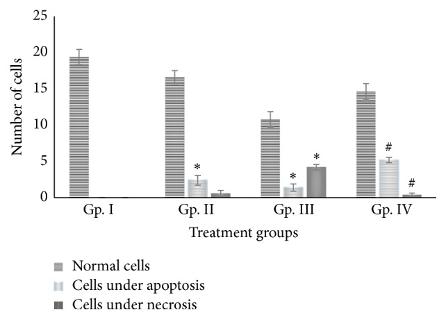
Cis-Diamminedichloroplatinum II- (CP-) induced neurotoxicity is one of the least explored aspects of this drug. Dorsal root ganglia (DRG) cells are considered as the primary target, and their damage plays a vital role in pathogenesis and etiology of CP-induced neurotoxicity. The present study is aimed at confirming if riboflavin (RF) has any protective role in shielding the DRG from CP-induced toxicity. After conducting the established treatment strategy on mice under photoillumination, it was observed that, despite the fact that RF alone is partially toxic, its combination with CP significantly ameliorated the drug-induced damage in DRG cells as evidenced by histological analysis. In addition, it was interesting to observe that the combination group (RF + CP) was able to induce apoptosis in the target cells up to a significant extent which is considered as the most preferred way of countering cancer cells. Therefore, RF can act as an effective adjuvant compound in CP-based chemoradiotherapy to improve clinical outcomes in the contemporary anticancer treatment regimes.
Figures

Similar articles
-
Cisplatin-induced neurotoxicity in vivo can be alleviated by riboflavin under photoillumination.Cancer Biother Radiopharm. 2013 Mar;28(2):160-8. doi: 10.1089/cbr.2012.1312. Epub 2012 Dec 7. Cancer Biother Radiopharm. 2013. PMID: 23215961
-
Inhibition of Ku70 in a high-glucose environment aggravates bupivacaine-induced dorsal root ganglion neurotoxicity.Toxicol Lett. 2020 Jan;318:104-113. doi: 10.1016/j.toxlet.2019.10.020. Epub 2019 Oct 28. Toxicol Lett. 2020. PMID: 31672611
-
Role of platinum DNA damage-induced transcriptional inhibition in chemotherapy-induced neuronal atrophy and peripheral neurotoxicity.J Neurochem. 2015 Dec;135(6):1099-112. doi: 10.1111/jnc.13355. Epub 2015 Oct 28. J Neurochem. 2015. PMID: 26364854
-
Vitamin B₂: a promising adjuvant in cisplatin based chemoradiotherapy by cellular redox management.Food Chem Toxicol. 2013 Sep;59:715-23. doi: 10.1016/j.fct.2013.07.018. Epub 2013 Jul 17. Food Chem Toxicol. 2013. PMID: 23872133 Review.
-
Chemotherapy-induced peripheral neuropathy: What do we know about mechanisms?Neurosci Lett. 2015 Jun 2;596:90-107. doi: 10.1016/j.neulet.2014.10.014. Epub 2014 Oct 22. Neurosci Lett. 2015. PMID: 25459280 Review.
Cited by
-
Pharmacological Effects of Cisplatin Combination with Natural Products in Cancer Chemotherapy.Int J Mol Sci. 2022 Jan 28;23(3):1532. doi: 10.3390/ijms23031532. Int J Mol Sci. 2022. PMID: 35163459 Free PMC article. Review.
-
ROS mediated antibacterial activity of photoilluminated riboflavin: A photodynamic mechanism against nosocomial infections.Toxicol Rep. 2019 Jan 9;6:136-142. doi: 10.1016/j.toxrep.2019.01.003. eCollection 2019. Toxicol Rep. 2019. PMID: 30671349 Free PMC article.
-
Nanoparticles for cancer therapy: a review of influencing factors and evaluation methods for biosafety.Clin Transl Oncol. 2023 Jul;25(7):2043-2055. doi: 10.1007/s12094-023-03117-5. Epub 2023 Feb 19. Clin Transl Oncol. 2023. PMID: 36807057 Review.
-
Antioxidant Supplementation in the Treatment of Neurotoxicity Induced by Platinum-Based Chemotherapeutics-A Review.Int J Mol Sci. 2020 Oct 20;21(20):7753. doi: 10.3390/ijms21207753. Int J Mol Sci. 2020. PMID: 33092125 Free PMC article. Review.
-
Riboflavin Inhibits Histamine-Dependent Itch by Modulating Transient Receptor Potential Vanilloid 1 (TRPV1).Front Mol Neurosci. 2021 Jun 18;14:643483. doi: 10.3389/fnmol.2021.643483. eCollection 2021. Front Mol Neurosci. 2021. PMID: 34220447 Free PMC article.
References
Publication types
MeSH terms
Substances
LinkOut - more resources
Full Text Sources
Other Literature Sources
Miscellaneous
