. 2016 Aug:8:59-67.
doi: 10.1016/j.redox.2015.12.011. Epub 2015 Dec 30.

Cellular and subcellular oxidative stress parameters following severe spinal cord injury

Affiliations

Cellular and subcellular oxidative stress parameters following severe spinal cord injury

Nishant P Visavadiya et al. Redox Biol. 2016 Aug.

Abstract

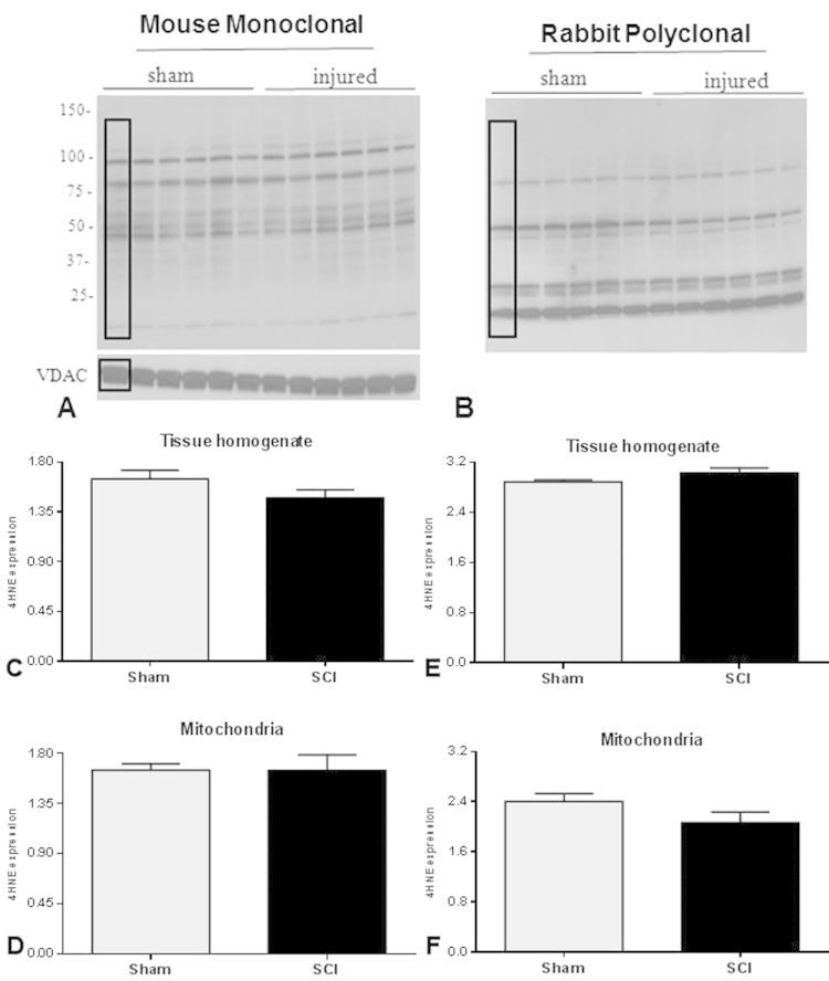
The present study undertook a comprehensive assessment of the acute biochemical oxidative stress parameters in both cellular and, notably, mitochondrial isolates following severe upper lumbar contusion spinal cord injury (SCI) in adult female Sprague Dawley rats. At 24h post-injury, spinal cord tissue homogenate and mitochondrial fractions were isolated concurrently and assessed for glutathione (GSH) content and production of nitric oxide (NO(•)), in addition to the presence of oxidative stress markers 3-nitrotyrosine (3-NT), protein carbonyl (PC), 4-hydroxynonenal (4-HNE) and lipid peroxidation (LPO). Moreover, we assessed production of superoxide (O2(•-)) and hydrogen peroxide (H2O2) in mitochondrial fractions. Quantitative biochemical analyses showed that compared to sham, SCI significantly lowered GSH content accompanied by increased NO(•) production in both cellular and mitochondrial fractions. SCI also resulted in increased O2(•-) and H2O2 levels in mitochondrial fractions. Western blot analysis further showed that reactive oxygen/nitrogen species (ROS/RNS) mediated PC and 3-NT production were significantly higher in both fractions after SCI. Conversely, neither 4-HNE levels nor LPO formation were increased at 24h after injury in either tissue homogenate or mitochondrial fractions. These results indicate that by 24h post-injury ROS-induced protein oxidation is more prominent compared to lipid oxidation, indicating a critical temporal distinction in secondary pathophysiology that is critical in designing therapeutic approaches to mitigate consequences of oxidative stress.

Keywords: 3-Nitrotyrosine; 4-Hydroxynonenal; Mitochondria; Protein carbonyl; RNS; ROS.


Figures

fx1
Graphical abstract
Fig. 1.
Fig. 1
GSH content was measured using the fluorescent probe MCB with GST reaction and was found to be significantly decreased 24 h after SCI in spinal cord tissue homogenate (A) and mitochondria (B) compared to Shams. Bars represent group means±SEM, n=6/group. *p<0.05 and **p<0.01.
Fig. 2.
Fig. 2
NO levels were assessed by standard Griess reagent and vanadium (III) chloride–based reduction assay. Spinal cord tissue homogenate (A) and mitochondria (B) showed significantly increased NO levels 24 h following SCI compared to sham. Bars represent group means±SEM, n=6/group. **p<0.01.
Fig. 3.
Fig. 3
Western blot images demonstrate 3-NT bands in tissue homogenate (A) and mitochondrial (B) fractions. Quantitative density values of 3-NT protein bands relative to internal controls from tissue homogenate (C) and mitochondrial fractions (D) showed that 24 h after SCI, 3-NT formation was significantly increased in both fractions compared to Shams. The boxes in A and B represent regions of interest used for comparative analyses. β-actin or VDAC were used as loading controls for tissue homogenate and mitochondria, respectively. Bars represent group means±SEM, n=6/group. *p<0.05.
Fig. 4.
Fig. 4
Panels A and B represent western blot images for PC banding patterns in tissue homogenate and mitochondrial fractions, respectively. Quantitative results showed that after 24 h, SCI significantly increased PC formation in tissue homogenate (C) and mitochondrial fractions (D). The arrows in panels A and B indicate specific BSA bands of ~66 kDa that underwent carbonylation. The boxes in A and B represent regions of interest used for density calculations. As loading controls, transferred membranes were stained with Coomassie Blue and relative densities of PC levels for each sample were normalized to total protein loaded. Bars represent group means±SEM, n=6/group. *p<0.05; ***p<0.001.
Fig. 5.
Fig. 5
Representative western blots demonstrate 4-HNE adducts formation using mouse monoclonal (A) and rabbit polyclonal (B) antibodies for mitochondrial fractions, using VDAC as a loading control. The bar graphs show that there were no changes in 4-HNE levels 24 h after SCI compared to Sham using either antibody in tissue homogenate (C and E) or mitochondrial (D and H) fractions. It should be noted that the PVDF membrane for mitochondria was stripped prior to re-probe with VDAC and then again for rabbit polyclonal antibody; hence a common VDAC band was used for normalization of both 4-HNE antibodies. The boxes in A and B represent regions of interest used for density calculations. Bars represent group means±SEM, n=6/group.
Fig. 6.
Fig. 6
LPO levels that were measured by TBA–TCA reagent were not different in either tissue homogenate (A) or mitochondrial (B) fractions 24 h following contusion SCI compared to Shams. Bars represent group means±SEM, n=6/group.
Fig. 7.
Fig. 7
Production of superoxide radical (O2-) in mitochondria was measured using DCF/HRP fluorescence dye. O2- production was significantly increased in mitochondrial fractions (A) at 24 h after SCI compared with Shams. Production of hydrogen peroxide (H2O2) in mitochondria was measured using Amplex Red/HRP fluorescence reagent and was also found to be significantly increased 24 h after SCI (B) compared to Shams. Bars represent group means±SEM, n=6/group. **p<0.01; ***p<0.001.

References

    1. Bains M., Hall E.D. Antioxidant therapies in traumatic brain and spinal cord injury. Biochim. Biophys. Acta. 2012;1822:675–684. - PMC - PubMed
    1. Fatima G., Sharma V.P., Das S.K., Mahdi A.A. Oxidative stress and antioxidative parameters in patients with spinal cord injury: implications in the pathogenesis of disease. Spinal Cord. 2015;53:3–6. - PubMed
    1. Hall E.D., Springer J.E. Neuroprotection and acute spinal cord injury: a reappraisal. NeuroRx: J. Am. Soc. Exp. Neurother. 2004;1:80–100. - PMC - PubMed
    1. McEwen M.L., Sullivan P.G., Rabchevsky A.G., Springer J.E. Targeting mitochondrial function for the treatment of acute spinal cord injury. Neurother.: J. Am. Soc. Exp. Neurother. 2011;8:168–179. - PMC - PubMed
    1. Rabchevsky A.G., Patel S.P., Springer J.E. Pharmacological interventions for spinal cord injury: where do we stand? How might we step forward? Pharmacol. Ther. 2011;132:15–29. - PubMed

Publication types