Review
. 2016 Jan 8;17(1):74.
doi: 10.3390/ijms17010074.

The Roles of RNase-L in Antimicrobial Immunity and the Cytoskeleton-Associated Innate Response

Affiliations
Review

The Roles of RNase-L in Antimicrobial Immunity and the Cytoskeleton-Associated Innate Response

Heather J Ezelle et al. Int J Mol Sci. .

Abstract

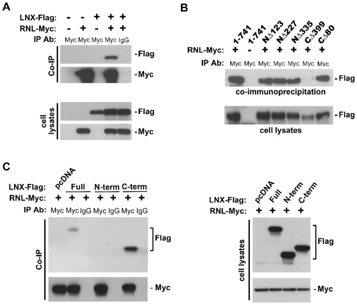
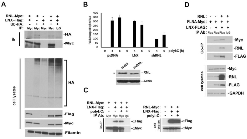
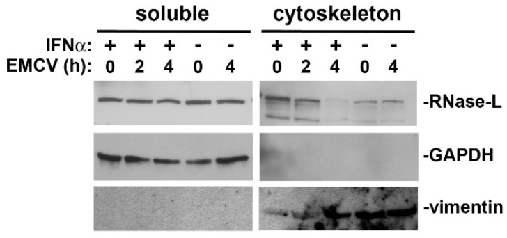
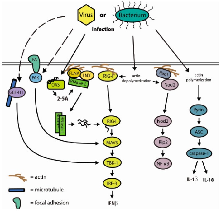
The interferon (IFN)-regulated endoribonuclease RNase-L is involved in multiple aspects of the antimicrobial innate immune response. It is the terminal component of an RNA cleavage pathway in which dsRNA induces the production of RNase-L-activating 2-5A by the 2'-5'-oligoadenylate synthetase. The active nuclease then cleaves ssRNAs, both cellular and viral, leading to downregulation of their expression and the generation of small RNAs capable of activating retinoic acid-inducible gene-I (RIG-I)-like receptors or the nucleotide-binding oligomerization domain-like receptor 3 (NLRP3) inflammasome. This leads to IFNβ expression and IL-1β activation respectively, in addition to broader effects on immune cell function. RNase-L is also one of a growing number of innate immune components that interact with the cell cytoskeleton. It can bind to several cytoskeletal proteins, including filamin A, an actin-binding protein that collaborates with RNase-L to maintain the cellular barrier to viral entry. This antiviral activity is independent of catalytic function, a unique mechanism for RNase-L. We also describe here the interaction of RNase-L with the E3 ubiquitin ligase and scaffolding protein, ligand of nump protein X (LNX), a regulator of tight junction proteins. In order to better understand the significance and context of these novel binding partners in the antimicrobial response, other innate immune protein interactions with the cytoskeleton are also discussed.

Keywords: LNX; RNase-L; actin; cytoskeleton; filamin A; inflammasome; innate immunity; interferon.


Figures

Figure 1
Figure 1
The 2′-5′-linked oligoadenylate (2-5A) System. IFN treatment induces the transcription of oligoadenylate synthetase (OAS), and virus infection produces dsRNA that activates OAS to synthesize 2-5A. Latent RNase-L binds 2-5A and oligomerizes into an active complex capable of cleaving ssRNA into retinoic acid-inducible gene-I (RIG-I) and nucleotide-binding oligomerization domain-like receptor 3 (NLRP3) inflammasome-activating small RNAs. Cleavage of target RNAs can regulate cellular and viral gene expression. RNase-L activation can be attenuated by phosphatase and 2′-phosphodiesterase mediated degradation of 2-5A. Solid arrows denote direct interactions and dashed arrows signify that intermediary steps occur.
Figure 2
Figure 2
RNase-L interacts with LNX. (A) 293T cells were transfected with pCMV3-RNL-myc, pcDNA3-hLNX-Flag, or the respective empty vectors using Lipofectamine 2000 (Invitrogen). Cell lysates were immunoprecipitated (IP) using anti-Myc-tag or IgG control antibodies bound to protein A/G agarose beads. Bound protein complexes were detected by Western blot analysis using the antibodies as indicated; (B) RNase-L and (C) LNX deletions were used in IP studies as in (A) to determine their respective interaction domains [43,127]. RNase-L deletions were subcloned from the pGEX4T3 plasmid (kindly provided by R.H. Silverman) into the eukaryotic expression plasmid pCMV3B using the BamHI and XhoI restriction sites.
Figure 3
Figure 3
Characterization and consequences of LNX expression. (A) Ubiquitylation of RNase-L is not enhanced by LNX expression. 293T cells were transfected with LNX, RNase-L, and HA-tagged ubiquitin (Ub). The presence of Ub-HA-conjugated RNase-L was determined by Western blot; (B) Hela cells were transfected with the indicated vectors. After 48 h, cells were transfected with polyI:C for 4 and 6 h and then analyzed by RT-qPCR for IFNβ expression, relative to rpl13a mRNA. Error bars represent standard deviation of two experiments. Western blot demonstrates shRNA knockdown of RNase-L expression. * indicates a non-specific band (C) 293T cells were transfected with LNX and RNase-L. After 48 h, cells were transfected with polyI:C for 6 h and then interaction was analyzed by IP; (D) Analysis of FLNA-myc and LNX-Flag interaction. FLNA and LNX were transfected into 293T cells in the presence or absence of RNase-L. Cell lysates were immunoprecipitated for LNX-Flag and Western blots probed for FLNA interaction. The presence of exogenous RNase-L did not alter the interaction.
Figure 4
Figure 4
Analysis of RNase-L association with the cytoskeleton. Caco-2 cells were treated with 500 U/mL hIFNα for 24 h and then infected with encephalomyocarditis virus (EMCV; MOI = 10) for 2 and 4 h. Soluble and cytoskeletal protein fractions were collected using the ProteoExtract Cytoskeleton Enrichment and Isolation Kit (Millipore) according to the manufacturer’s protocol. GAPDH and vimentin were used to evaluate soluble and cytoskeleton fraction purity, respectively.
Figure 5
Figure 5
Model of cytoskeletal interactions with components of innate immunity. Microbial infection creates perturbations in cytoskeletal interactions or actin polymerization that function as sensors to activate the innate immune response. The IFNβ and inflammasome pathways are key targets. Solid arrows denote direct interactions and dashed arrows signify that intermediary steps occur.

References

    1. Brand S., Zitzmann K., Dambacher J., Beigel F., Olszak T., Vlotides G., Eichhorst S.T., Goke B., Diepolder H., Auernhammer C.J. SOCS-1 inhibits expression of the antiviral proteins 2′,5′-OAS and MxA induced by the novel interferon-λs IL-28a and IL-29. Biochem. Biophys. Res. Commun. 2005;331:543–548. doi: 10.1016/j.bbrc.2005.04.004. - DOI - PubMed
    1. Der S.D., Zhou A., Williams B.R., Silverman R.H. Identification of genes differentially regulated by interferon α,β, or γ using oligonucleotide arrays. Proc. Natl. Acad. Sci. USA. 1998;95:15623–15628. doi: 10.1073/pnas.95.26.15623. - DOI - PMC - PubMed
    1. Hovanessian A.G. On the discovery of interferon-inducible, double-stranded RNA activated enzymes: The 2′-5′oligoadenylate synthetases and the protein kinase PKR. Cytokine Growth Factor Rev. 2007;18:351–361. doi: 10.1016/j.cytogfr.2007.06.003. - DOI - PubMed
    1. Tanaka N., Nakanishi M., Kusakabe Y., Goto Y., Kitade Y., Nakamura K.T. Structural basis for recognition of 2′,5′-linked oligoadenylates by human ribonuclease L. EMBO J. 2004;23:3929–3938. doi: 10.1038/sj.emboj.7600420. - DOI - PMC - PubMed
    1. Baglioni C., Minks M.A., Maroney P.A. Interferon action may be mediated by activation of a nuclease by pppa2′p5′A2′p5′A. Nature. 1978;273:684–687. doi: 10.1038/273684a0. - DOI - PubMed

Publication types

Substances

LinkOut - more resources