

The Importance of Thrombin in Cerebral Injury and Disease
- PMID: 26761005
- PMCID: PMC4730327
- DOI: 10.3390/ijms17010084
The Importance of Thrombin in Cerebral Injury and Disease
Abstract
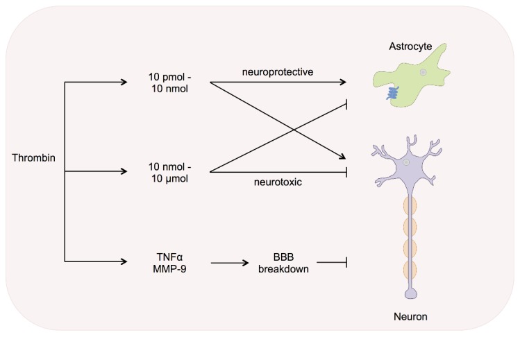
There is increasing evidence that prothrombin and its active derivative thrombin are expressed locally in the central nervous system. So far, little is known about the physiological and pathophysiological functions exerted by thrombin in the human brain. Extra-hepatic prothrombin expression has been identified in neuronal cells and astrocytes via mRNA measurement. The actual amount of brain derived prothrombin is expected to be 1% or less compared to that in the liver. The role in brain injury depends upon its concentration, as higher amounts cause neuroinflammation and apoptosis, while lower concentrations might even be cytoprotective. Its involvement in numerous diseases like Alzheimer's, multiple sclerosis, cerebral ischemia and haemorrhage is becoming increasingly clear. This review focuses on elucidation of the cerebral thrombin expression, local generation and its role in injury and disease of the central nervous system.
Keywords: Alzheimer’s disease; Parkinson’s disease; cerebral thrombin system; intracerebral haemorrhage; multiple sclerosis; prothrombin; stroke; thrombin.
Figures
Similar articles
-
Role of Thrombin in Central Nervous System Injury and Disease.Biomolecules. 2021 Apr 12;11(4):562. doi: 10.3390/biom11040562. Biomolecules. 2021. PMID: 33921354 Free PMC article. Review.
-
Prothrombin/thrombin and the thrombin receptors PAR-1 and PAR-4 in the brain: localization, expression and participation in neurodegenerative diseases.Thromb Haemost. 2008 Oct;100(4):576-81. Thromb Haemost. 2008. PMID: 18841278 Review.
-
Increased thrombin activity following reperfusion after ischemic stroke alters synaptic transmission in the hippocampus.J Neurochem. 2015 Dec;135(6):1140-8. doi: 10.1111/jnc.13372. Epub 2015 Oct 21. J Neurochem. 2015. PMID: 26390857
-
The involvement of thrombin in the pathogenesis of glioblastoma.J Neurosci Res. 2017 Oct;95(10):2080-2085. doi: 10.1002/jnr.24049. Epub 2017 Mar 19. J Neurosci Res. 2017. PMID: 28316070 Review.
-
Protease-activated receptors in neuronal development, neurodegeneration, and neuroprotection: thrombin as signaling molecule in the brain.Neuroscientist. 2004 Dec;10(6):501-12. doi: 10.1177/1073858404269955. Neuroscientist. 2004. PMID: 15534036 Review.
Cited by
-
Thrombin Aggravates Hypoxia/Reoxygenation Injury of Cardiomyocytes by Activating an Autophagy Pathway-Mediated by SIRT1.Med Sci Monit. 2021 May 1;27:e928480. doi: 10.12659/MSM.928480. Med Sci Monit. 2021. PMID: 33931577 Free PMC article.
-
Thrombin as Key Mediator of Seizure Development Following Traumatic Brain Injury.Front Pharmacol. 2020 Jan 14;10:1532. doi: 10.3389/fphar.2019.01532. eCollection 2019. Front Pharmacol. 2020. PMID: 32009953 Free PMC article.
-
Role of Thrombosis in Neurodegenerative Diseases: An Intricate Mechanism of Neurovascular Complications.Mol Neurobiol. 2025 Apr;62(4):4802-4836. doi: 10.1007/s12035-024-04589-4. Epub 2024 Nov 1. Mol Neurobiol. 2025. PMID: 39482419 Review.
-
Protease-Activated Receptor-1 Supports Locomotor Recovery by Biased Agonist Activated Protein C after Contusive Spinal Cord Injury.PLoS One. 2017 Jan 25;12(1):e0170512. doi: 10.1371/journal.pone.0170512. eCollection 2017. PLoS One. 2017. PMID: 28122028 Free PMC article.
-
Effects of Thrombin on the Neurovascular Unit in Cerebral Ischemia.Cell Mol Neurobiol. 2022 May;42(4):973-984. doi: 10.1007/s10571-020-01019-6. Epub 2021 Jan 3. Cell Mol Neurobiol. 2022. PMID: 33392917 Free PMC article. Review.
References
Publication types
MeSH terms
Substances
LinkOut - more resources
Full Text Sources
Other Literature Sources
Medical
