Differences in regional homogeneity between patients with Crohn's disease with and without abdominal pain revealed by resting-state functional magnetic resonance imaging
- PMID: 26761381
- PMCID: PMC4969077
- DOI: 10.1097/j.pain.0000000000000479
Differences in regional homogeneity between patients with Crohn's disease with and without abdominal pain revealed by resting-state functional magnetic resonance imaging
Abstract
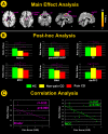
Abnormal pain processing in the central nervous system may be related to abdominal pain in patients with Crohn's disease (CD). The purpose of this study was to investigate changes in resting-state brain activity in patients with CD in remission and its relationship with the presence of abdominal pain. Twenty-five patients with CD and with abdominal pain, 25 patients with CD and without abdominal pain, and 32 healthy subjects were scanned using a 3.0-T functional magnetic resonance imaging scanner. Regional homogeneity (ReHo) was used to assess resting-state brain activity. Daily pain scores were collected 1 week before functional magnetic resonance imaging. We found that patients with abdominal pain exhibited lower ReHo values in the insula, middle cingulate cortex (MCC), and supplementary motor area and higher ReHo values in the temporal pole. In contrast, patients without abdominal pain exhibited lower ReHo values in the hippocampal/parahippocampal cortex and higher ReHo values in the dorsomedial prefrontal cortex (all P < 0.05, corrected). The ReHo values of the insula and MCC were significantly negatively correlated with daily pain scores for patients with abdominal pain (r = -0.53, P = 0.008 and r = -0.61, P = 0.002, respectively). These findings suggest that resting-state brain activities are different between remissive patients with CD with and without abdominal pain and that abnormal activities in insula and MCC are closely related to the severity of abdominal pain.
Figures


Similar articles
-
Alterations of Regional Homogeneity in Crohn's Disease With Psychological Disorders: A Resting-State fMRI Study.Front Neurol. 2022 Feb 2;13:817556. doi: 10.3389/fneur.2022.817556. eCollection 2022. Front Neurol. 2022. PMID: 35185768 Free PMC article.
-
Disruption of Periaqueductal Gray-default Mode Network Functional Connectivity in Patients with Crohn's Disease with Abdominal Pain.Neuroscience. 2023 May 1;517:96-104. doi: 10.1016/j.neuroscience.2023.03.002. Epub 2023 Mar 9. Neuroscience. 2023. PMID: 36898497
-
Abnormal regional homogeneity and its relationship with symptom severity in cervical dystonia: a rest state fMRI study.BMC Neurol. 2021 Feb 5;21(1):55. doi: 10.1186/s12883-021-02079-x. BMC Neurol. 2021. PMID: 33546628 Free PMC article.
-
Differences in brain gray matter volume in patients with Crohn's disease with and without abdominal pain.Oncotarget. 2017 Sep 22;8(55):93624-93632. doi: 10.18632/oncotarget.21161. eCollection 2017 Nov 7. Oncotarget. 2017. PMID: 29212177 Free PMC article.
-
Abnormal regional homogeneity in patients with irritable bowel syndrome: A resting-state functional MRI study.Neurogastroenterol Motil. 2015 Dec;27(12):1796-803. doi: 10.1111/nmo.12692. Epub 2015 Sep 25. Neurogastroenterol Motil. 2015. PMID: 26403620
Cited by
-
Altered functional connectivity of the amygdala in Crohn's disease.Brain Imaging Behav. 2020 Dec;14(6):2097-2106. doi: 10.1007/s11682-019-00159-8. Brain Imaging Behav. 2020. PMID: 31628591
-
Altered resting-state brain functional activities and networks in Crohn's disease: a systematic review.Front Neurosci. 2024 Jan 24;18:1319359. doi: 10.3389/fnins.2024.1319359. eCollection 2024. Front Neurosci. 2024. PMID: 38332859 Free PMC article.
-
Graph Theory Further Revealed Visual Spatial Working Memory Impairment in Patients with Inflammatory Bowel Disease.J Inflamm Res. 2024 May 8;17:2811-2823. doi: 10.2147/JIR.S462268. eCollection 2024. J Inflamm Res. 2024. PMID: 38737113 Free PMC article.
-
Wide-spread brain alterations early after the onset of Crohn's disease in children in remission-a pilot study.Front Neurosci. 2024 Dec 3;18:1491770. doi: 10.3389/fnins.2024.1491770. eCollection 2024. Front Neurosci. 2024. PMID: 39691628 Free PMC article.
-
Aberrant Brain Function in Active-Stage Ulcerative Colitis Patients: A Resting-State Functional MRI Study.Front Hum Neurosci. 2019 Apr 3;13:107. doi: 10.3389/fnhum.2019.00107. eCollection 2019. Front Hum Neurosci. 2019. PMID: 31001097 Free PMC article.
References
-
- Agostini A, Benuzzi F, Filippini N, Bertani A, Farinelli V, Marchetta C, Calabrese C, Rizzello F, Gionchetti P, Ercolani M, Campieri M, Nichelli P. New insights into the brain involvement in patients with Crohn's disease: a voxel-based morphometry study. Neurogastroenterol Motil. 2013;25:147–e82. - PubMed
-
- Agostini A, Filippini N, Benuzzi F, Bertani A, Scarcelli A, Leoni C, Farinelli V, Riso D, Tambasco R, Calabrese C, Rizzello F, Gionchetti P, Ercolani M, Nichelli P, Campieri M. Functional magnetic resonance imaging study reveals differences in the habituation to psychological stress in patients with Crohn's disease versus healthy controls. J Behav Med. 2013;36:477–87. - PubMed
-
- Agostini A, Filippini N, Cevolani D, Agati R, Leoni C, Tambasco R, Calabrese C, Rizzello F, Gionchetti P, Ercolani M, Leonardi M, Campieri M. Brain functional changes in patients with Ulcerative Colitis: an fMRI study on emotional processing. Inflamm Bowel Dis. 2011;17:1769–77. - PubMed
-
- Akbar A, Yiangou Y, Facer P, Brydon WG, Walters JR, Anand P, Ghosh S. Expression of the TRPV1 receptor differs in quiescent inflammatory bowel disease with or without abdominal pain. Gut. 2010;59:767–74. - PubMed
Publication types
MeSH terms
Substances
Grants and funding
LinkOut - more resources
Full Text Sources
Other Literature Sources
Medical

