

Review of Quantitative Ultrasound: Envelope Statistics and Backscatter Coefficient Imaging and Contributions to Diagnostic Ultrasound
- PMID: 26761606
- PMCID: PMC5551399
- DOI: 10.1109/TUFFC.2015.2513958
Review of Quantitative Ultrasound: Envelope Statistics and Backscatter Coefficient Imaging and Contributions to Diagnostic Ultrasound
Abstract
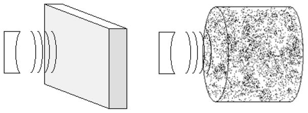
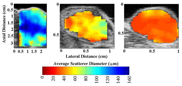
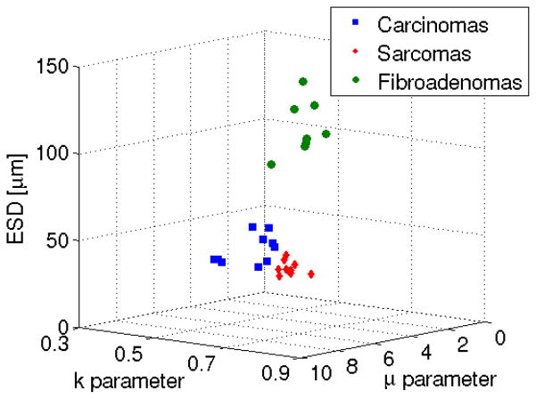
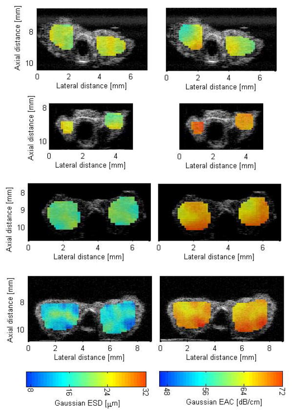
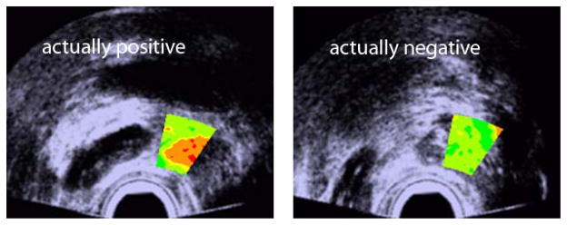
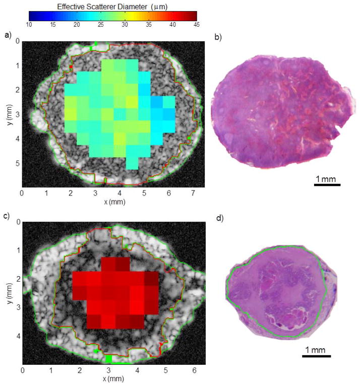
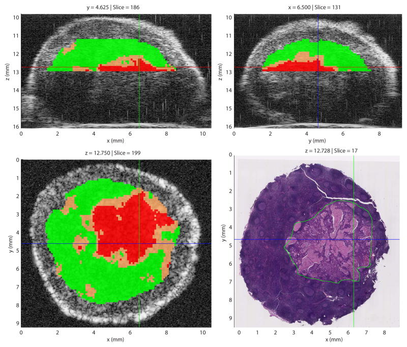
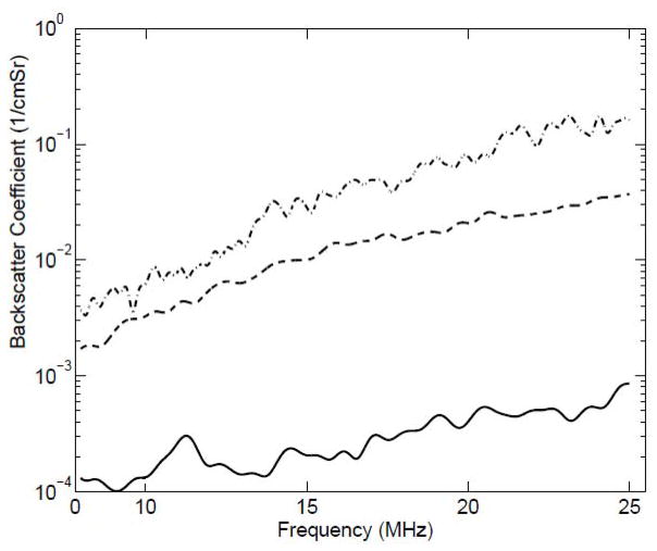
Conventional medical imaging technologies, including ultrasound, have continued to improve over the years. For example, in oncology, medical imaging is characterized by high sensitivity, i.e., the ability to detect anomalous tissue features, but the ability to classify these tissue features from images often lacks specificity. As a result, a large number of biopsies of tissues with suspicious image findings are performed each year with a vast majority of these biopsies resulting in a negative finding. To improve specificity of cancer imaging, quantitative imaging techniques can play an important role. Conventional ultrasound B-mode imaging is mainly qualitative in nature. However, quantitative ultrasound (QUS) imaging can provide specific numbers related to tissue features that can increase the specificity of image findings leading to improvements in diagnostic ultrasound. QUS imaging can encompass a wide variety of techniques including spectral-based parameterization, elastography, shear wave imaging, flow estimation, and envelope statistics. Currently, spectral-based parameterization and envelope statistics are not available on most conventional clinical ultrasound machines. However, in recent years, QUS techniques involving spectral-based parameterization and envelope statistics have demonstrated success in many applications, providing additional diagnostic capabilities. Spectral-based techniques include the estimation of the backscatter coefficient (BSC), estimation of attenuation, and estimation of scatterer properties such as the correlation length associated with an effective scatterer diameter (ESD) and the effective acoustic concentration (EAC) of scatterers. Envelope statistics include the estimation of the number density of scatterers and quantification of coherent to incoherent signals produced from the tissue. Challenges for clinical application include correctly accounting for attenuation effects and transmission losses and implementation of QUS on clinical devices. Successful clinical and preclinical applications demonstrating the ability of QUS to improve medical diagnostics include characterization of the myocardium during the cardiac cycle, cancer detection, classification of solid tumors and lymph nodes, detection and quantification of fatty liver disease, and monitoring and assessment of therapy.
Figures
References
-
- Spiegel PK. The first clinical X-ray made in America – 100 years. Amer J Roentgen. 1995;164:241–243. - PubMed
-
- Welch HG, William CB. Overdiagnosis in cancer. J Natl Cancer Inst. 2010;102:1–9. - PubMed
-
- Esserman L, Thompson I. Solving the overdiagnosis dilemma. J Natl Cancer Inst. 2010;102:582–583. - PubMed
-
- Silverstein M, Recht A, Lagois M, et al. Image-Detected Breast Cancer: State-of-the-Art Diagnosis and Treatment. J Am Coll Surg. 2009;209:504–520. - PubMed
-
- Oelze ML. Quantitative Ultrasound Techniques and Improvements to Diagnostic Ultrasonic Imaging. Proc IEEE Ultrasonics Symposium; Dresden, Germany. 2012. pp. 232–239.
Publication types
MeSH terms
Grants and funding
LinkOut - more resources
Full Text Sources
Other Literature Sources
Medical
Research Materials
Miscellaneous
