

Titanium Implant Impairment and Surrounding Muscle Cell Death Following High-Salt Diet: An In Vivo Study
- PMID: 26761710
- PMCID: PMC4711999
- DOI: 10.1371/journal.pone.0146873
Titanium Implant Impairment and Surrounding Muscle Cell Death Following High-Salt Diet: An In Vivo Study
Abstract
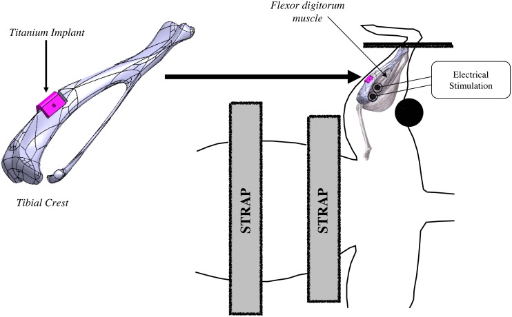
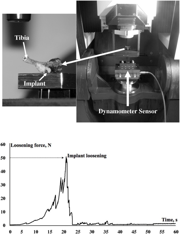
Aim of the study: High-salt consumption has been widely described as a risk factor for cardiovascular, renal and bone functions. In the present study, the extent to which high-salt diet could influence Ti6Al4V implant surface characteristic, its adhesion to rat tibial crest, and could modify muscle cell viability of two surrounding muscles, was investigated in vivo. These parameters have also been assessed following a NMES (neuro-myoelectrostimulation) program similar to that currently used in human care following arthroplasty.
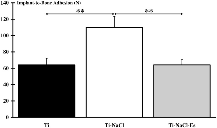
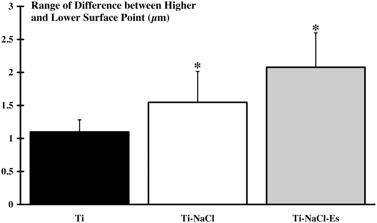
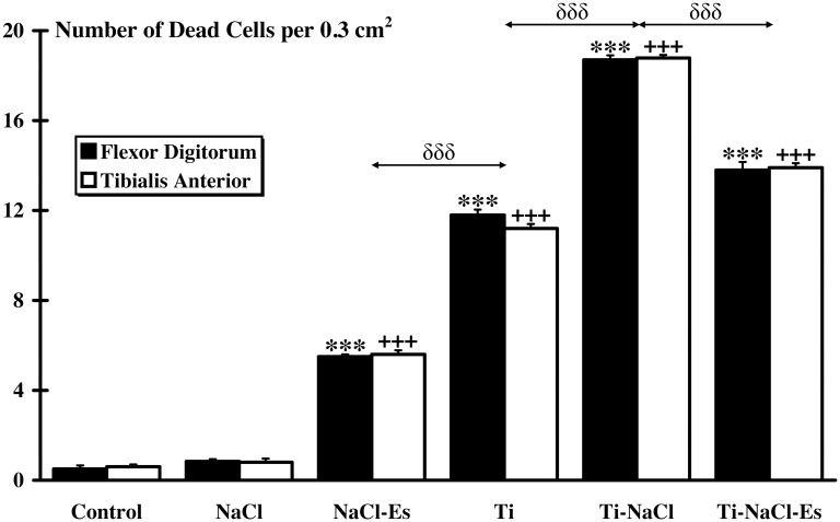
Results: After a three-week diet, a harmful effect on titanium implant surface and muscle cell viability was noted. This is probably due to salt corrosive effect on metal and then release of toxic substance around biologic tissue. Moreover, if the use of NMES with high-salt diet induced muscles damages, the latter were higher when implant was added. Unexpectedly, higher implant-to-bone adhesion was found for implanted animals receiving salt supplementation.
Conclusion: Our in vivo study highlights the potential dangerous effect of high-salt diet in arthroplasty based on titanium prosthesis. This effect appears to be more important when high-salt diet is combined with NMES.
Conflict of interest statement
Figures
Similar articles
-
Biocompatibility of Four Common Orthopedic Biomaterials Following a High-Salt Diet: An In Vivo Study.Int J Mol Sci. 2017 Jul 11;18(7):1489. doi: 10.3390/ijms18071489. Int J Mol Sci. 2017. PMID: 28696371 Free PMC article.
-
Titanium implant impairment and surrounding muscle cell death following neuro-myoelectrostimulation: An in vivo study.J Biomed Mater Res B Appl Biomater. 2015 Nov;103(8):1594-601. doi: 10.1002/jbm.b.33353. Epub 2014 Dec 23. J Biomed Mater Res B Appl Biomater. 2015. PMID: 25533414
-
Biocompatibility of four common orthopedic biomaterials following neuroelectromyostimulation: An in-vivo study.J Biomed Mater Res B Appl Biomater. 2018 Apr;106(3):1156-1164. doi: 10.1002/jbm.b.33927. Epub 2017 May 29. J Biomed Mater Res B Appl Biomater. 2018. PMID: 28556590
-
Titanium implant with nanostructured zirconia surface promotes maturation of peri-implant bone in osseointegration.Proc Inst Mech Eng H. 2013 May;227(5):510-22. doi: 10.1177/0954411913479300. Epub 2013 Mar 8. Proc Inst Mech Eng H. 2013. PMID: 23637261
-
Osseointegration of titanium, titanium alloy and zirconia dental implants: current knowledge and open questions.Periodontol 2000. 2017 Feb;73(1):22-40. doi: 10.1111/prd.12179. Periodontol 2000. 2017. PMID: 28000277 Review.
Cited by
-
Biocompatibility of Four Common Orthopedic Biomaterials Following a High-Salt Diet: An In Vivo Study.Int J Mol Sci. 2017 Jul 11;18(7):1489. doi: 10.3390/ijms18071489. Int J Mol Sci. 2017. PMID: 28696371 Free PMC article.
References
-
- Franco V, Oparil S. Salt sensitivity, a determinant of blood pressure, cardiovascular disease and survival. J Am Coll Nutr. 2006;25: 247S–255S. - PubMed
-
- Frassetto L, Morris RC Jr., Sellmeyer DE, Todd K, Sebastian A. Diet, evolution and aging—the pathophysiologic effects of the post-agricultural inversion of the potassium-to-sodium and base-to-chloride ratios in the human diet. Eur J Nutr. 2001;40: 200–213. - PubMed
-
- Frassetto LA, Morris RC Jr., Sellmeyer DE, Sebastian A. Adverse effects of sodium chloride on bone in the aging human population resulting from habitual consumption of typical American diets. J Nutr. 2008;138: 419S–422S. - PubMed
Publication types
MeSH terms
Substances
LinkOut - more resources
Full Text Sources
Other Literature Sources
Medical
Research Materials
