Review
. 2016 Jan 14:16:3.
doi: 10.1186/s12876-016-0419-2.

Pancreatic hamartoma: a case report and literature review

Affiliations
Review

Pancreatic hamartoma: a case report and literature review

Daisuke Matsushita et al. BMC Gastroenterol. .

Abstract

Background: Pancreatic hamartoma is an extremely rare benign disease of the pancreas. Only 30 cases have been reported to date.

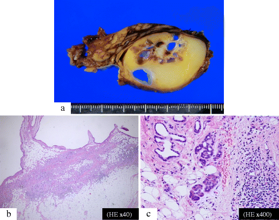
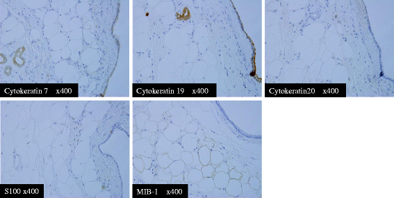
Case presentation: A 68-year-old man presented with an asymptomatic solid and multi-cystic lesion in the uncus of the pancreas, incidentally detected on abdominal enhanced computed tomography. The tumor was found to be a well-demarcated solid and multi-cystic lesion without any enhancement, measuring 4 cm in diameter. After 28 months of follow-up, the tumor enlarged. At 31 months after initial diagnosis, the patient underwent surgical resection because it was difficult to clinically determine whether the tumor was malignant or not. Macroscopically, the solid tumor consisted of yellow adipose tissue with a smooth thin capsule confined to the pancreatic uncus. The inner structure of the tumor consisted of multiple cysts with a white nodule between the cysts. Histologically, the solid part and the multi-cystic portion consisted of mature adipose tissue and colonization of dilated pancreatic ducts with mild fibrosis, respectively. Immunohistochemical findings revealed cytokeratin 7 and 19 positive staining in the epithelial cells of the ducts. Adipose tissue showed positive staining for S-100 protein and there were only a few MIB-1 positive cells. The tumor was then diagnosed as a pancreatic hamartoma.

Conclusion: Beside on the above findings, we suggest that the term "well-demarcated solid and cystic lesion with chronological morphological changes" could be a clinical keyword to describe pancreatic hamartomas.


Figures

Fig. 1
Fig. 1
Chronological changes seen on CT. a: First examination. The hypo-enhanced mass was 4.2 × 3.9 cm in size with solid and cystic lesions located in the uncus of the pancreas. b: At 21 months after first examination. The tumor shown is 3.9 × 3.6 cm in size. The cystic lesion (yellow arrow) had become smaller and the solid lesion (white arrow) had become larger. c: At 28 months after first examination. The tumor was 4.2 × 3.3 cm in size. The cystic lesion changed and displayed irregular margins
Fig. 2
Fig. 2
a, b, c. Chronological changes seen on MRCP. a: At the first examination. The tumor was observed as multiple cystic lesions. b: At 21 months after first examination. c: At 28 months after first examination. Figure 2-d, e, f, g. MRI appearance at 28 months after first examination. d: T1WI. A multi-cystic lesion with low intensity was surrounded by a mass with iso-intensity. e: T2WI. A multi-cystic lesion displays low- to iso-intensity. The surrounding tissue shows iso- to high intensity. f: T2-FAT-SAT. A multi-cystic high intensity lesion is shown. The surrounding tissue shows complete fat suppression. g: T2WI (coronal image). A ringed multi-cystic lesion with high intensity is surrounded by a smooth superficial mass. A small nodule is seen inside of the cyst ring
Fig. 3
Fig. 3
Intraoperative ultrasound examination. A well-demarcated honeycomb-like cystic lesion in the pancreatic uncus was found. There was no evidence of venous or ductal invasion
Fig. 4
Fig. 4
Macroscopic and pathological findings of the pancreatic hamartoma. a: Solid and cystic tumor in the pancreatic uncus with a smooth, thin capsule. Cysts were surrounded by yellow adipose tissue and a white nodule was found between the cysts. b: The solid lesion filled with mature adipose tissue. There was no evidence of mucinous products in the cysts and no evidence of malignancy in the epithelial cells of the cysts. c: The cystic lesion consisted of dilated ducts. Fibrosis and infiltration of monocytes were observed around the cysts. There were a few normal acini in the solid lesion, and the nodule located between the cysts consisted of stromal fibrosis without atypical cells
Fig. 5
Fig. 5
Immunohistochemical stains. The epithelial cells of the ducts expressed cytokeratin 7 and cytokeratin 19, but were negative for cytokeratin 20. The adipose tissue was positive for S-100 protein and mostly negative for MIB-1

References

    1. Volkan Adsay AN. Cystic lesions of the pancreas. Mod Pathol. 2007;20:S71–S93. doi: 10.1038/modpathol.3800706. - DOI - PubMed
    1. Raman SP, Hruban RH, Cameron JL, Wolfgang CL, Fishman EK. Pancreatic imaging mimics: part 2, pancreatic neuroendocrine tumors and their mimics. AJR Am J Roentgenol. 2012;199:309–318. doi: 10.2214/AJR.12.8627. - DOI - PubMed
    1. Anthony PP, Faber RG, Russell RC. Pseudotumours of the pancreas. Br Med J. 1977;1:814. doi: 10.1136/bmj.1.6064.814. - DOI - PMC - PubMed
    1. Burt TB, Condon VR, Matlak ME. Fetal pancreatic hamartoma. Pediatr Radiol. 1983;13:287–289. doi: 10.1007/BF00973350. - DOI - PubMed
    1. Flaherty MJ, Benjamin DR. Multicystic pancreatic hamartoma: a distinctive lesion with immunohistochemical and ultrastructural study. Hum Pathol. 1992;23:1309–1312. doi: 10.1016/0046-8177(92)90301-I. - DOI - PubMed