. 2016 Jan 14;529(7585):226-30.
doi: 10.1038/nature16527.

Intestinal epithelial tuft cells initiate type 2 mucosal immunity to helminth parasites

Affiliations

Intestinal epithelial tuft cells initiate type 2 mucosal immunity to helminth parasites

François Gerbe et al. Nature. .

Abstract

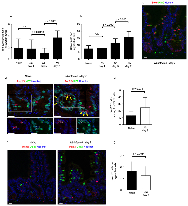
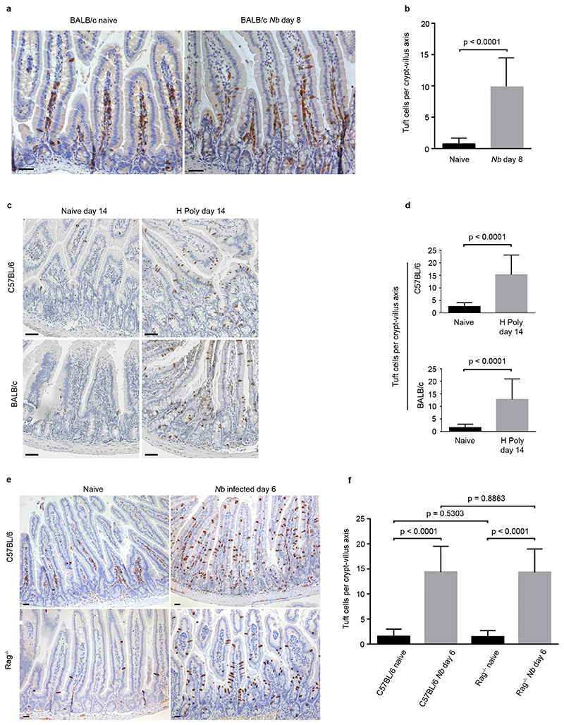
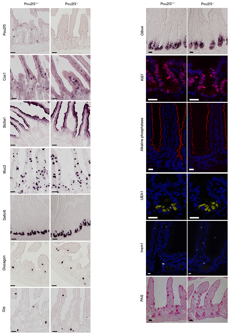
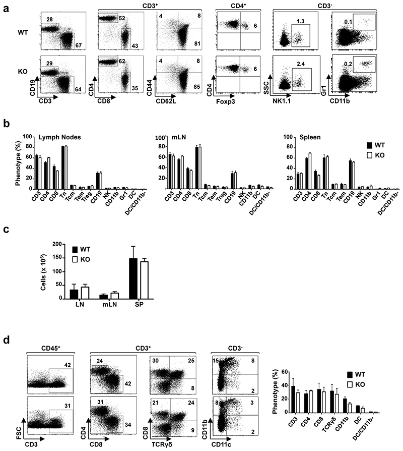
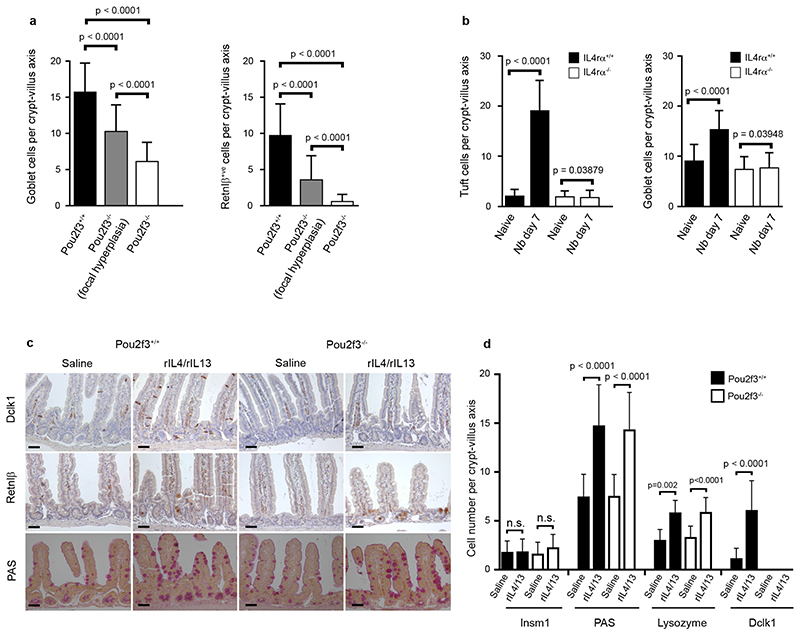
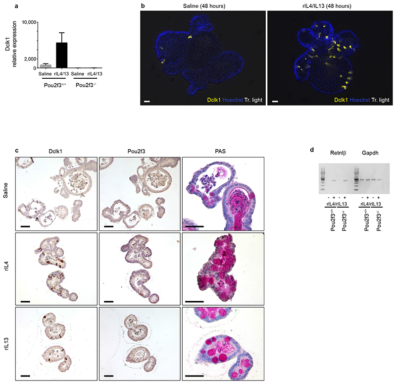
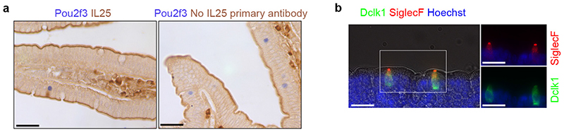
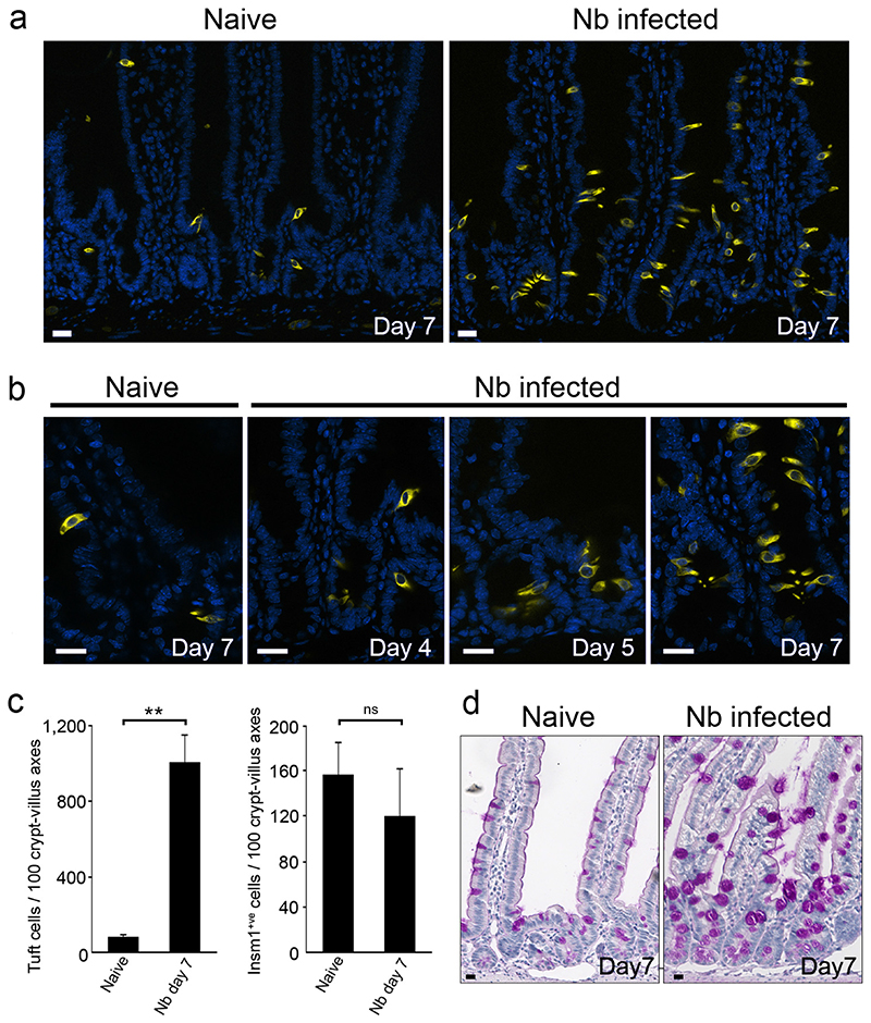
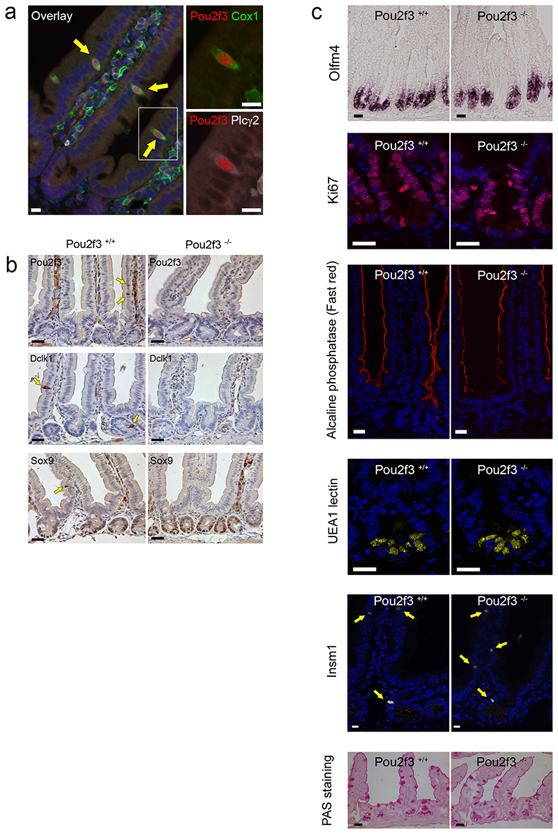
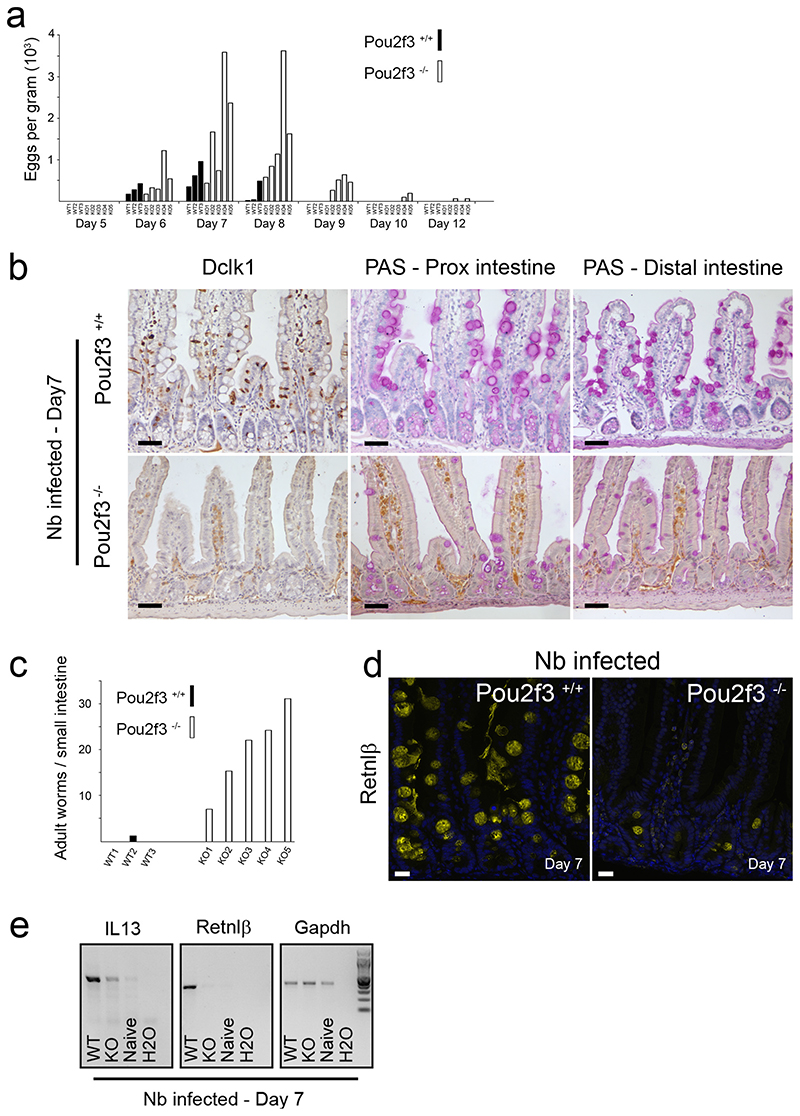
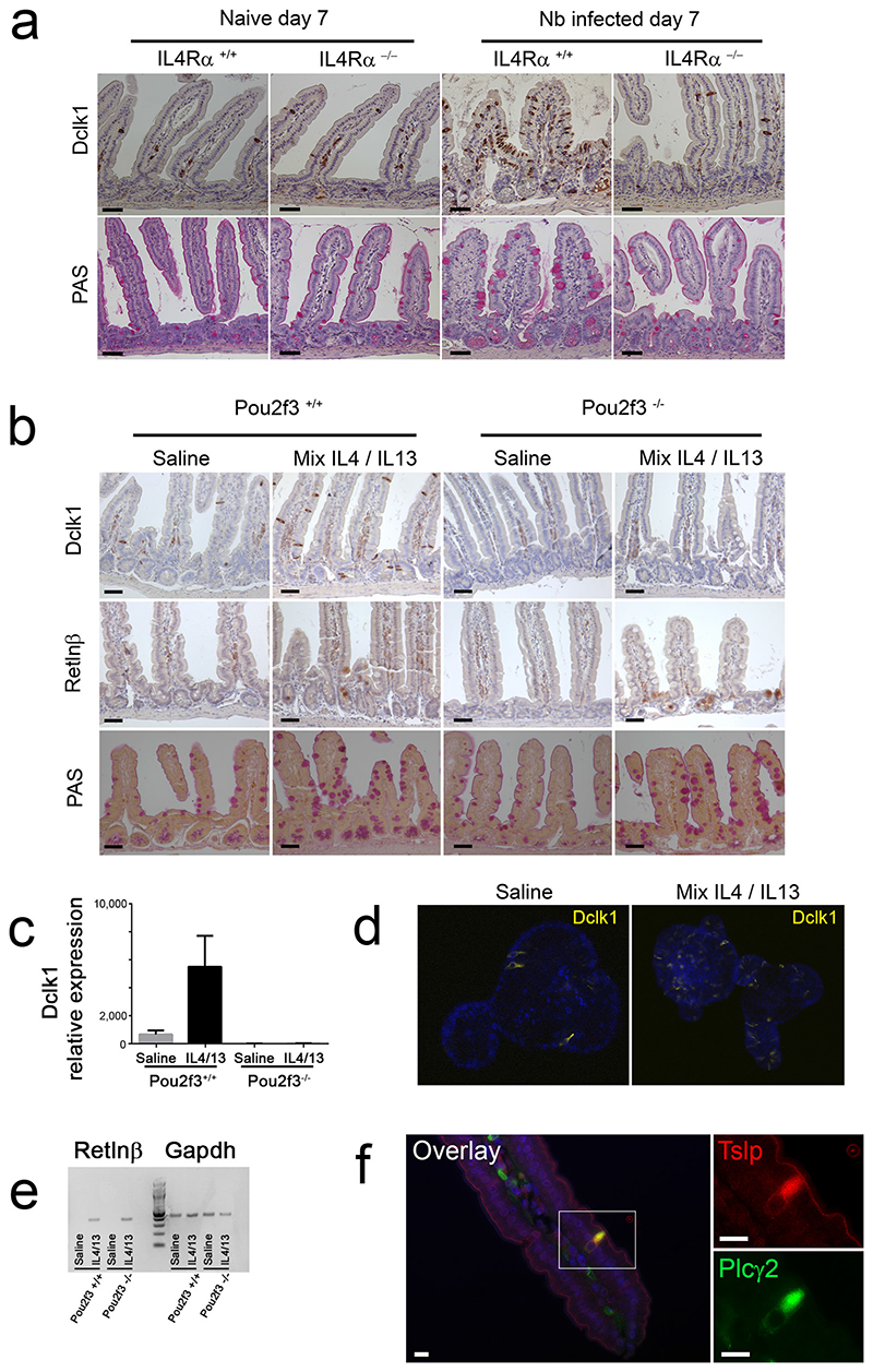
Helminth parasitic infections are a major global health and social burden. The host defence against helminths such as Nippostrongylus brasiliensis is orchestrated by type 2 cell-mediated immunity. Induction of type 2 cytokines, including interleukins (IL) IL-4 and IL-13, induce goblet cell hyperplasia with mucus production, ultimately resulting in worm expulsion. However, the mechanisms underlying the initiation of type 2 responses remain incompletely understood. Here we show that tuft cells, a rare epithelial cell type in the steady-state intestinal epithelium, are responsible for initiating type 2 responses to parasites by a cytokine-mediated cellular relay. Tuft cells have a Th2-related gene expression signature and we demonstrate that they undergo a rapid and extensive IL-4Rα-dependent amplification following infection with helminth parasites, owing to direct differentiation of epithelial crypt progenitor cells. We find that the Pou2f3 gene is essential for tuft cell specification. Pou2f3(-/-) mice lack intestinal tuft cells and have defective mucosal type 2 responses to helminth infection; goblet cell hyperplasia is abrogated and worm expulsion is compromised. Notably, IL-4Rα signalling is sufficient to induce expansion of the tuft cell lineage, and ectopic stimulation of this signalling cascade obviates the need for tuft cells in the epithelial cell remodelling of the intestine. Moreover, tuft cells secrete IL-25, thereby regulating type 2 immune responses. Our data reveal a novel function of intestinal epithelial tuft cells and demonstrate a cellular relay required for initiating mucosal type 2 immunity to helminth infection.


Conflict of interest statement

Data deposition: not applicable

Reprints and permissions information is available at www.nature.com/reprints

The authors declare no competing financial interest.

Figures

Extended Data Figure 1
Extended Data Figure 1. Epithelial differentiation parameters during Nb infection.
a, Graph showing the distribution of Dclk1+ tuft cells in naive and infected mice 4, 5 and 7 days post infection. Cells were counted in the crypt and villus compartments of n = 50 crypt–villus units per mouse with 3 mice per condition. Means of villus/crypt ratio of tuft cell numbers are shown. b, Quantification of the goblet cell hyperplasia in naive and infected mice 4, 5 and 7 days post infection (n = 50 crypt–villus units per mouse; 3 mice per condition). c, Neo-differentiating tuft cells following Nb infection are indistinguishable from the tuft cells found in naive mouse intestinal epithelium as shown with Sox9 and Plcγ2 stainings (n = 3 mice). d, Proliferation status of Pou2f3+ tuft cells in naive and infected mice, shown with co-expression with the Ki67 proliferation marker. Arrows indicate Ki67+ cells located at various positions along the crypt axis. e, Increased proliferation of Pou2f3+ tuft cells during response to Nb infection (n = 3 naive and 3 infected mice). f, Dclk1+ tuft cells and Insm1+ enteroendocrine cells are distinct populations (n = 3 mice). g, Decrease of the Insm1+ enteroendocrine cell population during type 2 responses to Nb infection, concomitant to the expansion of the tuft cell lineage 7 days post infection (n = 3 naive and 3 infected mice). All the histograms show means ± s.d. A two-tailed Student’s t-test with Welch’s correction was used, except for g where the 2 groups displayed comparable variances. All stainings were repeated 3 times.
Extended Data Figure 2
Extended Data Figure 2. Expansion of the tuft cell lineage is a common adaptation of the intestinal epithelium following infection with helminth parasites.
Tuft cell lineage expansion was assessed by Dclk1 immunohistochemistry in 2 different genetic backgrounds following infection with two different helminths, at the indicated time points. a, b, Naive and Nb-infected BALB/c mice. c, d, Naive and H. polygyrus-infected C57BL/6 and BALB/c mice. e, f, Naive and Nb-infected C57BL/6 and Rag−/− mice. b, d, f, n = 50 crypt–villus units per mouse; 3 mice per condition. Data are shown as means ± s.d. and P values are indicated. A two-tailed Student’s t-test with Welch’s correction was used. Scale bars, 20 μm. All experiments displayed in this figure were repeated 3 times.
Extended Data Figure 3
Extended Data Figure 3. Pou2f3 deficiency results in the specific absence of tuft cells in the intestinal epithelium.
Characterization of the intestinal epithelium in Pou2f3-deficient mice as compared with wild-type littermate controls (n = 3 mice of each genotype). Left, in situ hybridization showing absence of tuft cells (Pou2f3, Cox1) in Pou2f3−/− mice, whereas enterocytes (Slc5a1), goblet cells (Muc2), Paneth cells (Defcr6) and enteroendocrine cells (glucagon, Gip) are unaffected. Right, representative pictures of the in situ hybridization (Olfm4) and immunohistochemistry experiments underlying the quantitative analysis provided in Fig. 2c showing that the stem cells (Olfm4), proliferative compartment (Ki67), and differentiated cell types: enterocytes (alkaline phosphatase), Paneth (UEA1), enteroendocrine (Insm1) and goblet (PAS staining) cells populations are unaffected in the Pou2f3−/− mice. All panels show representative pictures of experiments replicated 3 times in 3 different mice. Scale bars, 20 μm.
Extended Data Figure 4
Extended Data Figure 4. Immune cell homeostasis is not altered in Pou2f3−/− tuft-cell-deficient mice.
a, The repartition of immune cells in wild-type and Pou2f3−/− mice was monitored by flow cytometry. The presence of T (CD3+), B (CD19+), CD4+, CD8+, naive (Tn; CD3+ CD62L+CD44-), central memory (Tcm; CD3+CD62L+CD44+), effector memory (Tem; CD3+CD62L-CD44+), regulatory (Treg; CD4+Foxp3+), natural killer (NK; CD3-NK1.1+) and myeloid (CD11b+Gr1+) cells was assessed by staining with fluorochrome-tagged antibodies and representative dot plots are shown. The percentages of positively-stained cells are indicated. b, Quantification of the different immune cells in lymph nodes (LN), mesenteric lymph nodes (mLN) and spleens (SP) of wild-type and Pou2f3−/− mice are presented. Data are means ± s.d. (n = 3 mice per genotype). c, Total cells in LN, mLN and SP of wild-type and Pou2f3−/− mice are presented as means ± s.d. (n = 3 mice per genotype). d, Immune lineage cells in the lamina propria of wild-type and Pou2f3−/− mice were monitored by flow cytometry after tissue dissociation. The percentage of T cells was assessed within the CD45+ haematopoietic gate, CD4, CD8 and gamma-delta T cells (CD8+TCR-γδ+) within the CD3+ gate and myeloid cells within the CD3- gate, as indicated. Representative dot plots are presented (left). Quantification of immune cells within the lamina propria are shown as means ± s.d. (n = 3 mice per group). No significant differences were detected for all cell types between wild-type and Pou2f3−/− mice (P > 0.05). A two-tailed Student’s t-test was used.
Extended Data Figure 5
Extended Data Figure 5. Equivalent immune responsiveness of wild-type and Pou2f3−/− lymphocytes.
a, The level of IL-2 and interferon gamma (IFN-γ) production by Pou2f3+/+ and Pou2f3−/− CD4 and CD8 lymph node T cells was monitored directly after ex vivo isolation and representative histograms are presented (left). Quantification of cytokine secreting CD4 and CD8 T cells are presented as means ± s.d. (n = 3 per group; P > 0.05). b, CFSE-loaded T cells were activated with immobilized anti-CD3/anti-CD28 antibodies for 2 days and proliferation was monitored as a function of fluorescence dilution. Representative histograms for CD4 and CD8 T cells are shown. c, IFN-γ production in wild-type and Pou2f3−/− lymphocytes was assessed at day 6 post CD3/CD28 stimulation and representative plots for CD4 and CD8 T cells are presented. d, Splenocytes from wild-type and Pou2f3−/− mice were activated with LPS+IL-4 for 40 h and levels of secreted IgG, IgG2a, IgG2b and IgA were monitored by ELISA. Means ± s.d. are presented. e, Splenocytes were activated as above and levels of TNF-α, IFN-γ, MCP-1, IL-10, IL-6, and IL-12 were monitored by cytometric bead array. Means ± s.d. are presented. A two-tailed Student’s t-test was used.
Extended Data Figure 6
Extended Data Figure 6. Defective induction of type 2 immunity in Pou2f3−/− mice following helminth infection.
a, Flow cytometry gating strategy for analysis of the innate ILC2 subset is shown. ILC2s were assessed within the CD45+ haematopoietic subset as lineage-CD127+ cells expressing KLRG1, GATA-3, Sca-1 and CD25 cell surface markers. Numbers represent the percentages of boxed cells. The staining strategy was validated using mLN cells from ZAP-70−/− mice as this subset is present at relatively high levels in these immunodeficient mice. b, The presence of ILC2 cells in mLNs of naive Pou2f3+/+ (WT) and Pou2f3−/− (KO) mice was assessed using the gating strategy shown above. Representative data from WT (n = 8) and KO (n = 5) mice are presented. c, Representative plots of ILC2 cells in lamina propria of naive WT (n = 7) and KO (n = 5) mice are shown (top). Quantifications of ILC2 are presented as means ± s.d. d, WT and KO mice were infected with N. brasiliensis and the presence of ILC2 in mLNs was assessed 5 days post infection. Representative plots are shown (n = 6 mice per group). e, Quantification of ILC2 cells in mLN of naive versus infected WT and KO mice. The percentage of ILC2s within the live gate (left) and the absolute numbers of ILC2s (right) are presented. Data are means ± s.d. (n = 5 for WT, n = 8 for KO, n = 6 for both groups of infected mice). **P = 0.01. f, The fold-increase in ILC2 (lineage-CD127+KLRG1+GATA-3+) and Th2 (CD3+CD4+Gata-3+) cells in mLN was assessed as a function of infection (n = 6 per group). The mean fold-increase ± s.d. in WT and KO mice is presented. *P = 0.02, ***P = 0.0005. A two-tailed Student’s t-test was used.
Extended Data Figure 7
Extended Data Figure 7. Signalling via IL-4Rα is required and sufficient to induce goblet and tuft cell hyperplasia.
a, Quantification of goblet (PAS and Retnlβ staining) cells in Pou2f3+/+ and Pou2f3−/− mice infected with Nb (day 7 post infection). In Pou2f3−/− mice, crypt–villus axes from both focally responding regions and the rest of the tissue were counted. b, Quantification of tuft cells (Dclk1 staining) and goblet cell hyperplasia (PAS) in Il4rα+/+ and Il4rα−/− mice. c, Histological analysis showing tuft (Dclk1 staining) and goblet (PAS and Retnlβ staining) cells in Pou2f3+/+ and Pou2f3−/− mice following treatment with a mixture of rIL-4 and rIL-13 for 5 days. n = 3 mice per condition. All panels show representative experiments replicated 3 times. Scale bars, 20 μm. d, Quantitative analysis of the changes in the different cell types of the intestinal epithelium of Pou2f3+/+ and Pou2f3−/− mice following treatment with a mixture of rIL-4 and rIL-13 during 5 days. For a, b, d, n = 50 crypt–villus axes counted in 3 mice per genotype or condition. Data are shown as means ± s.d. and P values are indicated. A two-tailed Student’s t-test with Welch’s correction was used.
Extended Data Figure 8
Extended Data Figure 8. Signalling via IL-4Rα is sufficient to induce goblet and tuft cell hyperplasia in mouse intestinal organoids.
a, Quantification of Dclkl expression analysis by qRT–PCR in Pou2f3+/+ and Pou2f3−/− organoids following rIL-4/rIL-13 treatment for 48 h to assess the presence and amplification of tuft cells. Means ± s.d., relative to Gapdh and Hprt, are presented. b, Expansion of the tuft cell lineage in wild-type organoids following rIL-4/rIL-13 administration (48 h) was monitored by Dclk1 staining. c, Expansion of the tuft cell lineage in wild-type organoids following IL-4 or IL-13 administration (48 h) was monitored by Dclk1, Pou2f3 and PAS stainings. Scale bars, 20 μm. d, Retnlβ expression in Pou2f3+/+ and Pou2f3−/− organoids was monitored as a function of rIL-4/rIL-13 treatment (48 h) by RT–PCR and data relative to Gapdh are presented. All panels show representative experiments from 3 independent organoid cultures, replicated 3 times.
Extended Data Figure 9
Extended Data Figure 9. Validation of the Siglec-F and IL-25 stainings.
a, Control experiment for specificity of the IL-25 immunohistochemistry, in presence (left) or absence (right) of IL-25 primary antibody. b, Immunohistochemistry showing specificity of Siglec-F as a marker for intestinal epithelial tuft cells. All panels show representative experiments from 3 independent mice, replicated 3 times.
Figure 1
Figure 1. Rapid amplification of the tuft cell lineage following infection with the helminth N. brasiliensis.
a : Presence of tuft cells in the intestinal epithelia of naïve and N. brasiliensis (Nb)-infected mice 7 days post infection, visualized by expression of the Dclk1 marker. b: 8.5 fold increase of tuft cells numbers (1,7±1,4 to 14,4±5,1 per cryptvillus axis) in Nb-infected mice as compared to naïve mice, 7 days post infection. Cells were counted in 50 crypt-villus units (n=3 mice per condition). Data are shown as means ± S.D. (p<0,0001). c: Changes in the tuft cell population in intestinal crypts are presented at the indicated time points post infection. Quantification is shown in Extended Data Figure 1a. d: Corresponding goblet cell hyperplasia associated with numerous and larger mucus vacuoles, detected by periodic acid-Schiff (PAS) staining. Dclk1 cells are also visualized in brown. Quantification is shown in Extended Data Figure 1b. For all panels, n=3 mice per condition. Bar: 20μm.
Figure 2
Figure 2. Absence of tuft cells in the intestinal epithelium of Pou2f3 transcription factorEdeficient mice.
a: Pou2f3 is expressed specifically in tuft cells of the intestinal epithelium as determined by co-staining for Pou2f3 and established markers of tuft cells such as Dclk1 and Gfi1b. b: Pou2f3 deletion results in the absence of tuft cells as monitored by staining intestinal epithelium from Pou2f3+/+ and Pou2f3−/− mice with Pou2f3-, Dclk1- and Sox9-specific antibodies. Bar: 20μm. c. Counting of 50 crypt-villus axes of 3 Pou2f3+/+ and 3 Pou2f3−/− mice revealed that the Pou2f3 deficiency does not affect the proliferation zone (p=0,76), stem cell compartment (p=0,53), enterocyte (not counted), goblet (p=0,95), Paneth (p=0,39) or enteroendocrine (p=0,12) cell lineages as monitored by Ki67, Olfm4, alkaline phosphatase, PAS staining, UEA1 lectin, and Insm1, respectively. Means ± S.D. and p values are indicated. For all panels, n=3 mice per condition.
Figure 3
Figure 3. Impaired type 2 responses in Pou2f3−/− tuft cell-deficient mice.
a: Live adult worm counts in the small intestines of Pou2f3+/+ and Pou2f3−/− mice at days 9 (left panel) and 13 (right panel) post infection with N. brasiliensis. Each bar represents an individual mouse and the WT or KO genotypes are indicated. b: Immunohistochemistry illustrating the proximal and distal small intestinal epithelium of infected Pou2f3+/+ and Pou2f3−/− mice 7 days after infection (3 mice per genotype). Tuft and goblet cells are revealed by Dclk1 and PAS staining, respectively, and the production of Resistin-like beta (Retnlβ) by goblet cells is analysed. Bar: 20μm. c: Quantification of IL13 and Retnlβ in the intestinal mucosa of naïve, and Nb-infected Pou2f3+/+ and Pou2f3−/− mice by RT-PCR, 7 days after infection. Representative gels are shown with relative Gapdh expression presented as an internal control. d: Histological analysis showing tuft (Dclk1 staining) and goblet (PAS staining) cells in naïve and Nb-infected Il4Rα+/+ and Il4Rα−/− mice 7 days post infection. For all panels, n>3 mice per condition. Bar: 20μm.
Figure 4
Figure 4. Tuft cells express IL25, and rIL25 is sufficient to initiate type 2 mucosal responses in the absence of tuft cells.
a: Analysis of Il25 mRNA expression in Pou2f3+/+ and Pou2f3−/− mice infected with N. brasiliensis, 9 days posbinfection, by RT-PCR. Data relative to Gapdh are presented. b: Immunohistochemistry showing IL25 expression in naïve and N. brasiliensis-infected wild type mice. Blue staining: nuclear Pou2f3 expression revealed with NBT/BCIP. Brown staining: IL25 expression revealed with DAB. n=3 naïve mice and 3 infected mice. Bar: 20μm. c: RT-PCR showing predominant IL25 and Pou2f3 mRNA expression in the tuft cells FACS-enriched fractions obtained from 3 independent mice. Gapdh is shown as a control for loading and RNA integrity. d: Rescue of the Pou2f3 deficiency by treatment with exogenous rIL25, as assessed by egg counts during a time course of infection with N. brasiliensis. N=6 mice per genotype and treatment condition. Means ± S.D. are presented, as well as p values. e: Scheme illustrating the function of tuft cells in initiating type 2 responses following infection with intestinal helminths. Left part: normal epithelium undergoing infection with an helminth. Right part: tuft cell-dependent epithelial remodelling during type 2 responses.

Comment in

References

    1. Hotez PJ, et al. Helminth infections: the great neglected tropical diseases. J Clin Invest. 2008;118:1311–1321. - PMC - PubMed
    1. Allen JE, Maizels RM. Diversity and dialogue in immunity to helminths. Nat Rev Immunol. 2011;11:375–388. - PubMed
    1. Herbert DR, et al. Intestinal epithelial cell secretion of RELM-beta protects against gastrointestinal worm infection. J Exp Med. 2009;206:2947–2957. - PMC - PubMed
    1. McKenzie GJ, Bancroft A, Grencis RK, McKenzie AN. A distinct role for interleukin-13 in Th2-cell-mediated immune responses. Current biology : CB. 1998;8:339–342. - PubMed
    1. Gerbe F, et al. Distinct ATOH1 and Neurog3 requirements define tuft cells as a new secretory cell type in the intestial epithelium. The Journal of Cell Biology. 2011;192:767–780. - PMC - PubMed

Publication types

MeSH terms