

Changes in the disposition of substance P in the rostral ventromedial medulla after inflammatory injury in the rat
- PMID: 26762802
- PMCID: PMC4738059
- DOI: 10.1016/j.neuroscience.2015.12.054
Changes in the disposition of substance P in the rostral ventromedial medulla after inflammatory injury in the rat
Abstract
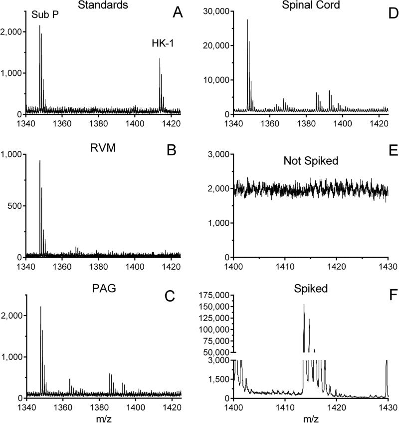
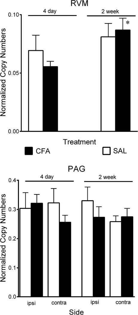
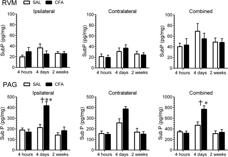
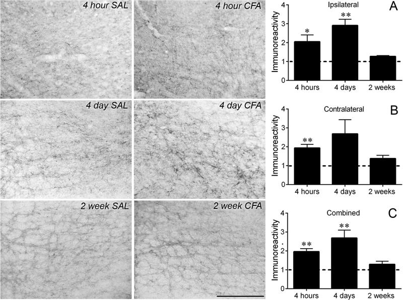
This study examined whether peripheral inflammatory injury increases the levels or changes the disposition of substance P (SubP) in the rostral ventromedial medulla (RVM), which serves as a central relay in bulbospinal pathways of pain modulation. Enzyme immunoassay and reverse transcriptase quantitative polymerase chain reaction were used to measure SubP protein and transcript, respectively, in tissue homogenates prepared from the RVM and the periaqueductal gray (PAG) and cuneiform nuclei of rats that had received an intraplantar injection of saline or complete Freund's adjuvant (CFA). Matrix-Assisted Laser Desorption/Ionization Time of Flight analysis confirmed that the RVM does not contain hemokinin-1 (HK-1), which can confound measurements of SubP because it is recognized equally well by commercial antibodies for SubP. Levels of SubP protein in the RVM were unchanged four hours, four days and two weeks after injection of CFA. Tac1 transcripts were similarly unchanged in the RVM four days or two weeks after CFA. In contrast, the density of SubP immunoreactive processes in the RVM increased 2-fold within four hours and 2.7-fold four days after CFA injection; it was unchanged at two weeks. SubP-immunoreactive processes in the RVM include axon terminals of neurons located in the PAG and cuneiform nucleus. SubP content in homogenates of the PAG and cuneiform nucleus was significantly increased four days after CFA, but not at four hours or two weeks. Tac1 transcripts in homogenates of these nuclei were unchanged four days and two weeks after CFA. These findings suggest that there is an increased mobilization of SubP within processes in the RVM shortly after injury accompanied by an increased synthesis of SubP in neurons that project to the RVM. These findings are consonant with the hypothesis that an increase in SubP release in the RVM contributes to the hyperalgesia that develops after peripheral inflammatory injury.
Keywords: hemokinin-1; hyperalgesia; inflammation; neurokinin-1 receptor; rostral ventromedial medulla; substance P.
Copyright © 2016 IBRO. Published by Elsevier Ltd. All rights reserved.
Figures
References
-
- Baranauskas G, Nistri A. Sensitization of pain pathways in the spinal cord: cellular mechanisms. Prog Neurobiol. 1998;54:349–365. - PubMed
-
- Beitz AJ. The nuclei of origin of brain stem enkephalin and substance P projections to the rodent nucleus raphe magnus. Neuroscience. 1982;7:2753–2768. - PubMed
-
- Berger A, Paige CJ. Hemokinin-1 has Substance P-like function in U-251 MG astrocytoma cells: a pharmacological and functional study. J Neuroimmunol. 2005;164:48–56. - PubMed
-
- Borbely E, Hajna Z, Sandor K, Kereskai L, Toth I, Pinter E, Nagy P, Szolcsanyi J, Quinn J, Zimmer A, Stewart J, Paige C, Berger A, Helyes Z. Role of tachykinin 1 and 4 gene-derived neuropeptides and the neurokinin 1 receptor in adjuvant-induced chronic arthritis of the mouse. PLoS One. 2013;8:e61684. - PMC - PubMed
Publication types
MeSH terms
Substances
Grants and funding
LinkOut - more resources
Full Text Sources
Other Literature Sources
Medical
Miscellaneous
