

Multiplexed neurochemical signaling by neurons of the ventral tegmental area
- PMID: 26763116
- PMCID: PMC4818729
- DOI: 10.1016/j.jchemneu.2015.12.016
Multiplexed neurochemical signaling by neurons of the ventral tegmental area
Abstract
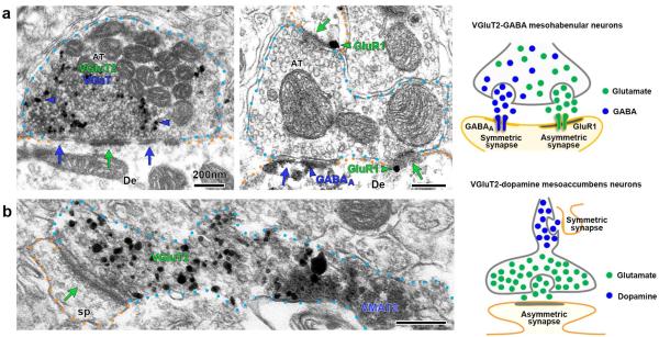
The ventral tegmental area (VTA) is an evolutionarily conserved structure that has roles in reward-seeking, safety-seeking, learning, motivation, and neuropsychiatric disorders such as addiction and depression. The involvement of the VTA in these various behaviors and disorders is paralleled by its diverse signaling mechanisms. Here we review recent advances in our understanding of neuronal diversity in the VTA with a focus on cell phenotypes that participate in 'multiplexed' neurotransmission involving distinct signaling mechanisms. First, we describe the cellular diversity within the VTA, including neurons capable of transmitting dopamine, glutamate or GABA as well as neurons capable of multiplexing combinations of these neurotransmitters. Next, we describe the complex synaptic architecture used by VTA neurons in order to accommodate the transmission of multiple transmitters. We specifically cover recent findings showing that VTA multiplexed neurotransmission may be mediated by either the segregation of dopamine and glutamate into distinct microdomains within a single axon or by the integration of glutamate and GABA into a single axon terminal. In addition, we discuss our current understanding of the functional role that these multiplexed signaling pathways have in the lateral habenula and the nucleus accumbens. Finally, we consider the putative roles of VTA multiplexed neurotransmission in synaptic plasticity and discuss how changes in VTA multiplexed neurons may relate to various psychopathologies including drug addiction and depression.
Keywords: Addiction; Aversion; Co-transmission; Depression; Dopamine; GABA; Glutamate; Reward.
Published by Elsevier B.V.
Figures

References
-
- Adcock RA, Thangavel A, Whitfield-Gabrieli S, Knutson B, Gabrieli JD. Reward-motivated learning: mesolimbic activation precedes memory formation. Neuron. 2006;50(3):507–517. - PubMed
-
- Aizawa H, Kobayashi M, Tanaka S, Fukai T, Okamoto H. Molecular characterization of the subnuclei in rat habenula. Journal of Comparative Neurology. 2012;520(18):4051–4066. - PubMed
-
- Alfahel-Kakunda A, Silverman WF. Calcium-binding proteins in the substantia nigra and ventral tegmental area during development: correlation with dopaminergic compartmentalization. Developmental brain research. 1997;103(1):9–20. - PubMed
Publication types
MeSH terms
Substances
Grants and funding
LinkOut - more resources
Full Text Sources
Other Literature Sources
