. 2016 Mar 4;291(10):5396-405.
doi: 10.1074/jbc.M115.713123. Epub 2016 Jan 13.

Loss of α-Tubulin Acetylation Is Associated with TGF-β-induced Epithelial-Mesenchymal Transition

Affiliations

Loss of α-Tubulin Acetylation Is Associated with TGF-β-induced Epithelial-Mesenchymal Transition

Shuchen Gu et al. J Biol Chem. .

Abstract

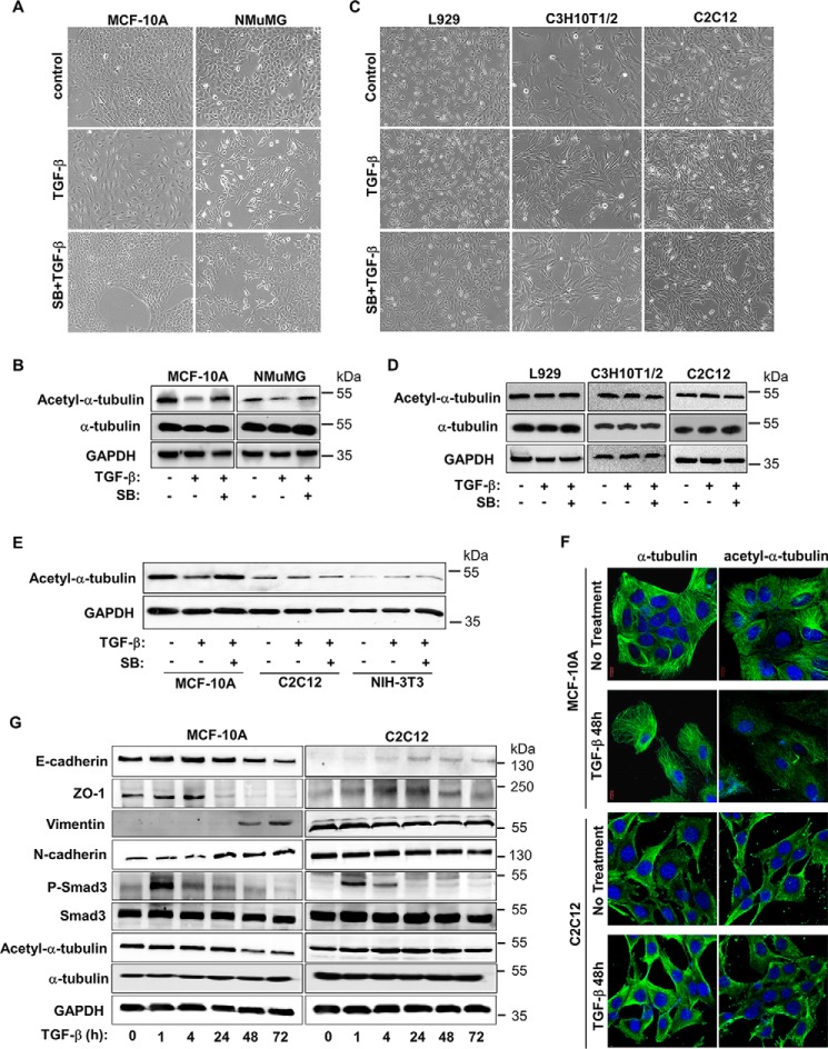
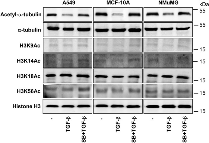
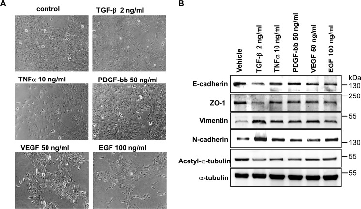
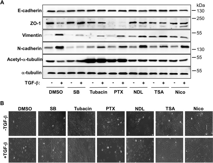
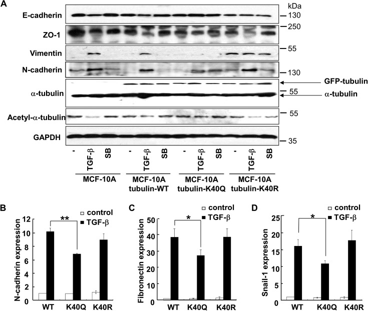
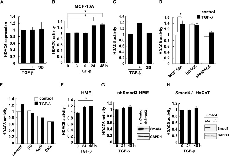
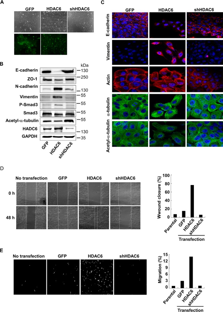
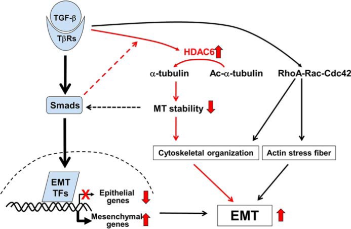
The epithelial-to-mesenchymal transition (EMT) is a process by which differentiated epithelial cells reprogram gene expression, lose their junctions and polarity, reorganize their cytoskeleton, increase cell motility and assume a mesenchymal morphology. Despite the critical functions of the microtubule (MT) in cytoskeletal organization, how it participates in EMT induction and maintenance remains poorly understood. Here we report that acetylated α-tubulin, which plays an important role in microtubule (MT) stabilization and cell morphology, can serve as a novel regulator and marker of EMT. A high level of acetylated α-tubulin was correlated with epithelial morphology and it profoundly decreased during TGF-β-induced EMT. We found that TGF-β increased the activity of HDAC6, a major deacetylase of α-tubulin, without affecting its expression levels. Treatment with HDAC6 inhibitor tubacin or TGF-β type I receptor inhibitor SB431542 restored the level of acetylated α-tubulin and consequently blocked EMT. Our results demonstrate that acetylated α-tubulin can serve as a marker of EMT and that HDAC6 represents an important regulator during EMT process.

Keywords: acetylation; epithelial-mesenchymal transition (EMT); histone deacetylase 6 (HDAC6); microtubule; transforming growth factor β (TGF-β).


Figures

FIGURE 1.
FIGURE 1.
Level of acetylated α-tubulin is decreased in TGF-β-induced EMT. A, TGF-β induces EMT in MCF-10A and NMuMG cells. Cells were treated with TGF-β (2 ng/ml) 48 h and/or SB431542 (5 μm), and then examined under microscope. B, TGF-β decreases the level of acetyl-α-tubulin in MCF-10A and NMuMG cells. Cells were treated with TGF-β (2 ng/ml) 48 h and/or SB431542 (5 μm), and then analyzed by using specific antibodies recognizing acetyl-α-tubulin, α-tubulin, and GAPDH. C, TGF-β exhibites no effect on morphology of L929, C3H10T1/2, and C2C12 cells. Cells were treated similarly as described in A. D, TGF-β does not decrease the level of acetyl-α-tubulin in L929, C3H10T1/2 and C2C12 cells. Cells were treated similarly as described in C and Western blotting as in B. E, TGF-β decreases the level of acetyl-α-tubulin in MCF-10A, but not C2C12 and NIH-3T3 cells. Immunoblotting analysis of acetyl-α-tubulin and GAPDH in MCF-10A, C2C12, and NIH-3T3 cells was done as described in B. F, TGF-β decreases the level of acetyl-α-tubulin in MCF-10A, but not C2C12 cells. Cells were treated with 2 ng/ml TGF-β (48 h), immunostained with specific antibodies for acetyl-α-tubulin or α-tubulin, and analyzed under confocal laser scanning microscopic as described under “Experimental Procedures.” Magnification 100×. G, immunoblotting analysis of E-cadherin, ZO-1, vimentin, N-cadherin, HDAC6, P-Smad3, Smad3, acetyl-α-tubulin, α-tubulin, and GAPDH. MCF-10A and C2C12 cells were stimulated with 2 ng/ml of TGF-β for the indicated time periods.
FIGURE 2.
FIGURE 2.
Global levels of Histone H3 acetylation are not decreased in TGF-β-induced EMT. MCF-10A, A549, and NMuMG cells were exposed to TGF-β (2 ng/ml, 48 h) in the presence of absence of SB431542 (5 μm, prior 2 h +additional 48 h). Acetyl-Histone H3 (lys9) or H3K9Ac, H3K14Ac, H3K18Ac, H3K56Ac, Histone H3, acetyl-α-tubulin, and GAPDH were examined by immunoblotting.
FIGURE 3.
FIGURE 3.
Level of acetylated α-tubulin is decreased in EMT induced by multiple growth factors. A, immunoblotting analysis of E-cadherin, ZO-1, Vimentin, N-cadherin, acetyl-α-tubulin, and α-tubulin in MCF-10A cells. Cells treated with TGF-β (2 ng/ml), or TNFα (10 ng/ml), PDGF-bb (50 ng/ml), VEGF (50 ng/ml) or (EGF 100 ng/ml). B, morphology of MCF-10A cells. Culture conditions were the same as those in A.
FIGURE 4.
FIGURE 4.
Inhibition of α-tubulin acetylation or disruption of its stability blocks EMT. A, immunoblotting analysis of E-cadherin, ZO-1, Vimentin, N-cadherin, acetyl-α-tubulin, and α-tubulin in MCF-10A cells. Cells were treated with SB431542 (5 μm), Tubacin (5 μm), Paclitaxol (PTX) (5 nm), Nocodazol (NDL) (5 nm), TSA (50 ng/liter), or Nicotiamide (Nico) (10 mm), followed by co-incubation with TGF-β (2 ng/ml). B, morphology of MCF-10A cells. Culture conditions were the same as those in A.
FIGURE 5.
FIGURE 5.
An acetylation-mimicking mutant of α-tubulin attenuates TGF-β-induced EMT. A, immunoblotting analysis of E-cadherin, ZO-1, Vimentin, N-cadherin, acetyl-α-tubulin, and α-tubulin in MCF-10A cells. MCF-10A cells were transfected to express wild type (WT), K40Q, or K40R mutant of α-tubulin. Cells were then treated with 2 ng/ml of TGF-β in the presence or absence of 5 μm of SB431542. GFP-tubulin and endogenous α-tubulin were indicated by arrows. B, K40Q mutant decreases TGF-β-induced expression of N-cadherin. MCF-10A cells infected with lentiviruses harboring α-tubulin WT, K40Q, or K40R were treated with TGF-β (2 ng/ml) 48 h. Total RNAs were isolated from treated cells and the N-cadherin mRNA levels were analyzed by using qRT-PCR. **, p < 0.01. C, K40Q mutant decreases TGF-β-induced expression of Fibronectin. qRT-PCR analysis was done as described in panel B. *, p < 0.05. D, K40Q mutant decreases TGF-β-induced expression of Snail-1. qRT-PCR analysis was done as described in panel B. *, p < 0.05.
FIGURE 6.
FIGURE 6.
TGF-β increases the activity of HDAC6. A, TGF-β and SB431542 have no effects on the mRNA levels of HDAC6 in MCF-10A cells. MCF-10A cells were treated with TGF-β (2 ng/ml) or SB431542 (5 μm) for 48 h. Total RNAs were isolated from treated cells and the HDAC6 mRNA levels in the treated cells were analyzed by using qRT-PCR. *, p < 0.05. B, TGF-β activates HDAC6 in MCF-10A cells. MCF-10A cells were treated with 2 ng/ml of TGF-β for the indicated time periods. Measurement of HDAC6 activity in cell lysates was carried out by using ELISA-based enzymatic assay as described under “Experimental Procedures.” C, TGF-β-induced HDAC6 activation is inhibited by SB431542 in MCF-10A cells. Measurement of HDAC6 activity was done as described in panel B. D, TGF-β activates HDAC6 in MCF-10A cells. MCF-10A cells stably expressing GFP (control), HDAC6 or shHDAC6 were treated with 2 ng/ml of TGF-β (48 h). Measurement of HDAC6 activity was done as described in panel B. E, TGF-β-induced HDAC6 activation is inhibited by SB431542, ActD and CHX in MCF-10A cells. MCF-10A cells were treated with SB431542, ActD and CHX for 48 h. HDAC6 activity was measured as described in panel B. F, TGF-β activates HDAC6 in HME cells. HME cells were treated with 2 ng/ml of TGF-β for the indicated time periods. HDAC6 activity was measured as described in panel B. G, TGF-β-induced HDAC6 activation is inhibited by Smad3 knockdown in HME cells. HME cells stably expressing an shRNA against Smad3 were treated with 2 ng/ml of TGF-β for the indicated time periods. HDAC6 activity was measured as described in panel B. Right, knockdown of Smad3 in HME was shown by Western blotting using indicated antibodies. H, TGF-β-induced HDAC6 activation is inhibited by Smad4 knock out in HaCaT cells. HaCaT cells with the Smad4 gene ablated were treated with 2 ng/ml of TGF-β for the indicated time periods. HDAC6 activity was measured as described in panel B. Right, knock-out of Smad4 in HaCaT was shown by Western blotting using indicated antibodies.
FIGURE 7.
FIGURE 7.
Increased expression of HDAC6 induces EMT. A, morphology of MCF-10A cells stably expressing GFP (control), HDAC6, or shHDAC6. Cells were examined under a fluorescence microscope. Top, bright field; bottom, GFP fluorescence. B, HDAC6 induces EMT marker expression. MCF-10A cells stably expressing GFP (control), HDAC6 or shHDAC6 were subject to immunoblotting analysis using antibodies against E-cadherin, ZO-1, vimentin, N-cadherin, HDAC6, P-Smad3, Smad3, acetyl-α-tubulin, and α-tubulin. C, decreased acetyl-α-tubulin is correlated with EMT marker gene expression. Confocal laser scanning microscopy was used to analyze the levels of E-cadherin, vimentin, actin, acetyl-α-tubulin, and α-tubulin. Magnification 100×. D, HDAC6 promotes cell motility. Confluent monolayer of MCF-10A cells were scratched with a pipette tip to create a cell-free area. 24 h after addition of TGF-β (2 ng/ml), the wound closure was recorded by microphotography of the same region (Left). Quantification of cell motility (Right) was done by measuring the distance between the invading front of cells in three random selected microscopic fields for each condition and time point. The degree of motility is expressed as percentage of wound closure as compared with untransfected cells. E, HDAC6 promotes cell invasiveness. MCF-10A cells were cultured in the Transwell culture chambers with an 8-μm pore-size polycarbonate filter according to the manufacturer's instructions. 24 h later, invaded cells that were attached to the lower surface of the filter were stained with DAPI. The slices were imaged (Left). Quantification (Right) was by counting cell number in the lower surface. The degree of invasiveness is expressed as percentage of total cells seeded as compared with untransfected cells.
FIGURE 8.
FIGURE 8.
Working Model for TGF-β-induced EMT. In addition to the transcriptional reprogramming during TGF-β-induced EMT, TGF-β induces HDAC6 activity that converts acetyl-α-tubulin into α-tubulin. This deacetylation promotes cytoskeletal reorganization that favors EMT.

References

    1. Hay E. D. (1995) An overview of epithelio-mesenchymal transformation. Acta Anatomica 154, 8–20 - PubMed
    1. Kalluri R., and Weinberg R. A. (2009) The basics of epithelial-mesenchymal transition. J. Clin. Invest. 119, 1420–1428 - PMC - PubMed
    1. Zavadil J., and Böttinger E. P. (2005) TGF-β and epithelial-to-mesenchymal transitions. Oncogene 24, 5764–5774 - PubMed
    1. Moustakas A., and Heldin C. H. (2007) Signaling networks guiding epithelial-mesenchymal transitions during embryogenesis and cancer progression. Cancer Science 98, 1512–1520 - PMC - PubMed
    1. Xu J., Lamouille S., and Derynck R. (2009) TGF-beta-induced epithelial to mesenchymal transition. Cell Res. 19, 156–172 - PMC - PubMed

Publication types

LinkOut - more resources