. 2016 Jan;136(1):34-44.
doi: 10.1038/JID.2015.354.

A Guide to Studying Human Hair Follicle Cycling In Vivo

Affiliations

A Guide to Studying Human Hair Follicle Cycling In Vivo

Ji Won Oh et al. J Invest Dermatol. 2016 Jan.

Erratum in

  • Erratum.
    [No authors listed] [No authors listed] J Invest Dermatol. 2016 Apr;136(4):883. doi: 10.1016/j.jid.2016.02.023. Epub 2016 Mar 21. J Invest Dermatol. 2016. PMID: 27140062 No abstract available.

Abstract

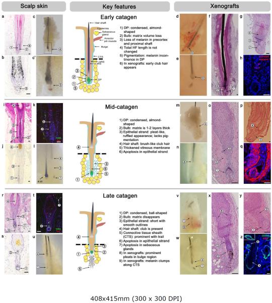
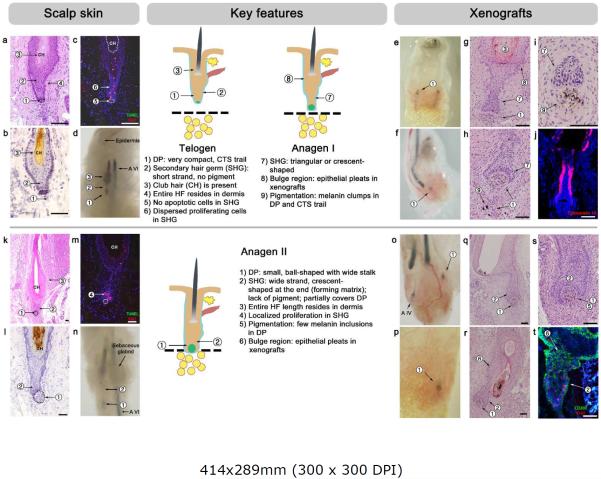
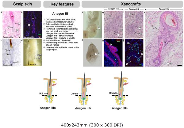
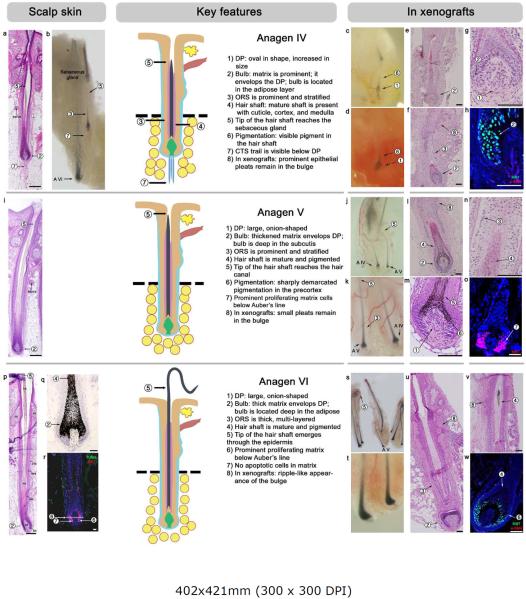
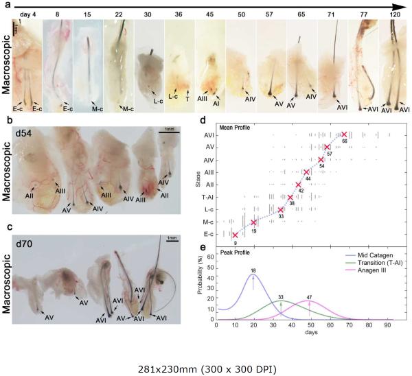
Hair follicles (HFs) undergo lifelong cyclical transformations, progressing through stages of rapid growth (anagen), regression (catagen), and relative "quiescence" (telogen). Given that HF cycling abnormalities underlie many human hair growth disorders, the accurate classification of individual cycle stages within skin biopsies is clinically important and essential for hair research. For preclinical human hair research purposes, human scalp skin can be xenografted onto immunocompromised mice to study human HF cycling and manipulate long-lasting anagen in vivo. Although available for mice, a comprehensive guide on how to recognize different human hair cycle stages in vivo is lacking. In this article, we present such a guide, which uses objective, well-defined, and reproducible criteria, and integrates simple morphological indicators with advanced, (immuno)-histochemical markers. This guide also characterizes human HF cycling in xenografts and highlights the utility of this model for in vivo hair research. Detailed schematic drawings and representative micrographs provide examples of how best to identify human HF stages, even in suboptimally sectioned tissue, and practical recommendations are given for designing human-on-mouse hair cycle experiments. Thus, this guide seeks to offer a benchmark for human hair cycle stage classification, for both hair research experts and newcomers to the field.


Figures

Figure 1
Figure 1. Catagen
For practical reasons, catagen was subdivided into three, easily recognizable stages: early, mid-, and late catagen. For each stage, a schematic drawing is provided, with key and auxiliary features numbered and marked. For HFs-IS, 5–10% of all HFs are in catagen. In xenografts, approximately three quarters of HFs-XG undergo “dystrophic catagen”, during which a club hair fails to form (see Supplementary Figure S1). (a–h) Early catagen in HFs-IS (a-c') and HFs-XG (d-h). Key features at this stage are matrix volume loss, loss of pigment at the proximal end of the hair shaft, melanin incontinence into the DP, and the appearance of apoptotic cells. On in situ, HF length remains unchanged compared to anagen VI HFs. In xenografts, the peak day for early catagen is post-grafting day 3. (i–q) Mid-catagen in HFs-IS (i-l) and HFs-XG (m-q). Key features at this stage are a shrinking matrix, which is only 1–2 cell layers thick, thin epithelial strand with pleated outlines and apoptotic cells, presence of the brush-like club hair, and a thick vitreous membrane. On in situ, the proximal portion of the HF is still within the adipose layer. In xenografts, the peak day for mid-catagen is post-grafting day 18. (r–z) Late catagen in HFs-IS (r-u) and HFs-XG (v-z). Key features at this stage are a smaller, ball-shaped DP, absence of the hair matrix, shortened (compared to mid-catagen) epithelial strand, prominent connective tissue sheath with the trail below the DP, melanin clumps in the trail, and ongoing apoptosis in the epithelial strand. Additionally, on in situ, apoptosis occurs in the sebaceous gland. In xenografts, the bulge region develops prominent pleats. This stage peaks on post-grafting day 29. Hosts: SCID mice (panels - e, g, j, m, q, x, y), nude mice (panels - d, f, h, n, o, p, v, w, z). Scale bars: 100 um.
Figure 2
Figure 2. Telogen and anagen I, II
For each stage, a schematic drawing is provided, and key and auxiliary features are numbered and marked. (a–d) Telogen. HFs with typical telogen morphology represent 1–2% of all HFs on in situ findings and are generally lacking in xenografts. Key features at this stage are a very small DP and a short secondary hair germ that lacks apoptotic cells. The entire length of the telogen HF rests in the dermis. (e–j) Anagen I. HFs with anagen I morphology are generally not found in situ but are common in xenografts, peaking on post-grafting day 33. Key features at this stage are a small DP, a secondary hair germ shaped as triangle or small crescent, and an initiation of proliferation at the base of the germ. (k–t) Anagen II in HFs-IS (k-n) and HFs-XG (o-t). Key features at this stage are a small DP with a wide stalk (compared to telogen and anagen I), an enlarged secondary hair germ with prominent crescent shape, and a localized proliferation hotspot at the base of the germ. On in situ, the entire length of the HF rests in the dermis. In xenografts, the peak day for anagen II is post-grafting day 40. Hosts: SCID mice (panels - f, g, h, j, o, q, r, s), nude mice (panels - e, i, p, t). Scale bars: 100 um.
Figure 3
Figure 3. Anagen III
Schematic drawings of HFs are provided, and key and auxiliary features are numbered and marked. Key features at this stage are an enlarged, oval-shaped DP (compared to anagen II), the presence of a newly formed, albeit small matrix (4–5 cell layers thick), small, but visible IRS, and a hair shaft that lacks pigmentation. On in situ, a newly formed hair bulb enters into the adipose layer. In xenografts, three anagen III sub-stages can be identified on the basis of combined IRS and hair shaft morphology. The bulge region of HFs-XG shows prominent pleats. This stage peaks on post-grafting day 47. Hosts: SCID mice (panels - f, h, j), nude mice (panels - e, g, i, k). Scale bars: 100 um.
Figure 4
Figure 4. Anagen IV, V and VI
For each stage, a schematic drawing is provided, and key and auxiliary features are numbered and marked. (a–h) Anagen IV in HFs-IS (a, b) and HFs-XG (c-h). Key features at this stage are a prominent matrix, stratified ORS, and a mature hair shaft that reaches the level of the sebaceous gland. On in situ, the hair bulb is in the adipose layer, but the connective tissue trail can still be seen (it becomes lost during anagen V). Xenografted anagen IV HFs show prominent pleats in the bulge region and peak on post-grafting day 60. (i–o) Anagen V in HFs-IS (i) and HFs-XG (j-o). Key features at this stage are a large, onion-shaped DP, significantly increased pigmentation (compared to anagen IV) with sharp demarcation at Auber's line, and a mature hair shaft that reaches the hair canal. On in situ, the connective tissue trail disappears (compared to anagen IV). Xenografted anagen V HFs maintain pleats in the bulge region and peak on post-grafting day 63. (p–w) Anagen VI in HFs-IS (p-r) and HFs-XG (s-w). 90–95% of all HFs-IS are in anagen VI, and all HFs-XG progress to anagen VI by post-grafting day 92. At this stage HFs achieve their maximum size, and the hair shaft tip extends far above the skin surface. There are no apoptotic cells compared to early catagen. Hosts: SCID mice (panels - e, f, g, m, n, s, t, u, w), nude mice (panels - c, d, h, j, k, l, o, v). Scale bars: 100 um.
Figure 5
Figure 5. Xenograft model optimization
(a) Representative gross morphology of human HFs-XG showing post-grafted hair cycle resetting dynamics: follicles progress through sequential catagen sub-stages, telogen-to-anagen transition stage, and then anagen sub-stages. (b, c) Representative images showing hair cycle heterogeneity of HFs-XG at day 54 (b) and 70 (c). (d) Comprehensive hair cycle staging of human HFs-XG during the first 90 days (x-axis). Average time point values for each sub-stage (y-axis) are shown with the average regression curve overlaid over the scatter plot of individual HFs stage values (assessed upon biopsy; each dot represents one biopsied HF). (e) Statistical analysis of the frequency at which the indicated stage appears (early catagen, telogen-to-anagen I transition, and anagen III). Arrows denote the post-grafting time with the greatest probability of selecting HFs-XG at the indicated stage based on the Naïve Bayes classifier analysis. Further details (for every hair cycle sub-stage) can be found on Supplementary Figure S5. Hosts: SCID mice (panels – a (days 15, 22, 50, 57, 65) and c), nude mice (panels – a (days 4, 8, 30, 36, 45, 71, 77, 120) and b). Scale bars: a, b, c – 1mm.

Comment in

References

    1. Al-Nuaimi Y, Hardman JA, Biro T, et al. A meeting of two chronobiological systems: circadian proteins Period1 and BMAL1 modulate the human hair cycle clock. The Journal of investigative dermatology. 2014;134:610–9. - PubMed
    1. Arck PC, Handjiski B, Kuhlmei A, et al. Mast cell deficient and neurokinin-1 receptor knockout mice are protected from stress-induced hair growth inhibition. J Mol Med (Berl) 2005;83:386–96. - PubMed
    1. Atanaskova Mesinkovska N, Bergfeld WF. Hair: what is new in diagnosis and management? Female pattern hair loss update: diagnosis and treatment. Dermatol Clin. 2013;31:119–27. - PubMed
    1. Bernard BA. The human hair follicle, a bistable organ? Experimental dermatology. 2012;21:401–3. - PubMed
    1. Bodo E, Tobin DJ, Kamenisch Y, et al. Dissecting the impact of chemotherapy on the human hair follicle: a pragmatic in vitro assay for studying the pathogenesis and potential management of hair follicle dystrophy. Am J Pathol. 2007;171:1153–67. - PMC - PubMed

Publication types