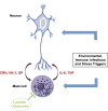
Neuropeptides CRH, SP, HK-1, and Inflammatory Cytokines IL-6 and TNF Are Increased in Serum of Patients with Fibromyalgia Syndrome, Implicating Mast Cells
- PMID: 26763911
- PMCID: PMC4767394
- DOI: 10.1124/jpet.115.230060
Neuropeptides CRH, SP, HK-1, and Inflammatory Cytokines IL-6 and TNF Are Increased in Serum of Patients with Fibromyalgia Syndrome, Implicating Mast Cells
Abstract
Fibromyalgia syndrome (FMS) is a chronic, idiopathic condition of widespread musculoskeletal pain affecting more women than men. Even though clinical studies have provided evidence of altered central pain pathways, the lack of definitive pathogenesis or reliable objective markers has hampered development of effective treatments. Here we report that the neuropeptides corticotropin-releasing hormone (CRH), substance P (SP), and SP-structurally-related hemokinin-1 (HK-1) were significantly (P = 0.026, P < 0.0001, and P = 0.002, respectively) elevated (0.82 ± 0.57 ng/ml, 0.39 ± 0.18 ng/ml, and 7.98 ± 3.12 ng/ml, respectively) in the serum of patients with FMS compared with healthy controls (0.49 ± 0.26 ng/ml, 0.12 ± 0.1 ng/ml, and 5.71 ± 1.08 ng/ml, respectively). Moreover, SP and HK-1 levels were positively correlated (Pearson r = 0.45, P = 0.002) in FMS. The serum concentrations of the inflammatory cytokines interleukin (IL)-6 and tumor necrosis factor (TNF) were also significantly (P = 0.029 and P = 0.006, respectively) higher (2.97 ± 2.35 pg/ml and 0.92 ± 0.31 pg/ml, respectively) in the FMS group compared with healthy subjects (1.79 ± 0.62 pg/ml and 0.69 ± 0.16 pg/ml, respectively). In contrast, serum IL-31 and IL-33 levels were significantly lower (P = 0.0001 and P = 0.044, respectively) in the FMS patients (849.5 ± 1005 pg/ml and 923.2 ± 1284 pg/ml, respectively) in comparison with healthy controls (1281 ± 806.4 pg/ml and 3149 ± 4073 pg/ml, respectively). FMS serum levels of neurotensin were not different from controls. We had previously shown that CRH and SP stimulate IL-6 and TNF release from mast cells (MCs). Our current results indicate that neuropeptides could stimulate MCs to secrete inflammatory cytokines that contribute importantly to the symptoms of FMS. Treatment directed at preventing the secretion or antagonizing these elevated neuroimmune markers, both centrally and peripherally, may prove to be useful in the management of FMS.
Copyright © 2016 by The American Society for Pharmacology and Experimental Therapeutics.
Figures


Similar articles
-
Effects of an Extract of Salmon Milt on Symptoms and Serum TNF and Substance P in Patients With Fibromyalgia Syndrome.Clin Ther. 2019 Aug;41(8):1564-1574.e2. doi: 10.1016/j.clinthera.2019.05.019. Epub 2019 Jul 11. Clin Ther. 2019. PMID: 31303280
-
Mast Cells, Neuroinflammation and Pain in Fibromyalgia Syndrome.Front Cell Neurosci. 2019 Aug 2;13:353. doi: 10.3389/fncel.2019.00353. eCollection 2019. Front Cell Neurosci. 2019. PMID: 31427928 Free PMC article. Review.
-
Elevated serum high-sensitivity C-reactive protein levels in fibromyalgia syndrome patients correlate with body mass index, interleukin-6, interleukin-8, erythrocyte sedimentation rate.Rheumatol Int. 2013 May;33(5):1259-64. doi: 10.1007/s00296-012-2538-6. Epub 2012 Nov 4. Rheumatol Int. 2013. PMID: 23124693
-
Evaluating relationship in cytokines level, Fibromyalgia Impact Questionnaire and Body Mass Index in women with Fibromyalgia syndrome.J Back Musculoskelet Rehabil. 2016;29(1):145-9. doi: 10.3233/BMR-150610. J Back Musculoskelet Rehabil. 2016. PMID: 26406191
-
Activation of Mast Cells by Neuropeptides: The Role of Pro-Inflammatory and Anti-Inflammatory Cytokines.Int J Mol Sci. 2023 Mar 2;24(5):4811. doi: 10.3390/ijms24054811. Int J Mol Sci. 2023. PMID: 36902240 Free PMC article. Review.
Cited by
-
Fibromyalgia: Pathogenesis, Mechanisms, Diagnosis and Treatment Options Update.Int J Mol Sci. 2021 Apr 9;22(8):3891. doi: 10.3390/ijms22083891. Int J Mol Sci. 2021. PMID: 33918736 Free PMC article. Review.
-
CRF serum levels differentiate PTSD from healthy controls and TBI in military veterans.Psychiatr Res Clin Pract. 2021 Winter;3(4):153-162. doi: 10.1176/appi.prcp.20210017. Epub 2021 Jun 1. Psychiatr Res Clin Pract. 2021. PMID: 35211666 Free PMC article.
-
Inflammation and Interferon Signatures in Peripheral B-Lymphocytes and Sera of Individuals With Fibromyalgia.Front Immunol. 2022 May 26;13:874490. doi: 10.3389/fimmu.2022.874490. eCollection 2022. Front Immunol. 2022. PMID: 35693781 Free PMC article.
-
The role of ultrasonographic synovial assessment in rheumatoid arthritis patients with concomitant fibromyalgia.Arch Rheumatol. 2022 Sep 20;38(2):174-182. doi: 10.46497/ArchRheumatol.2023.9585. eCollection 2023 Jun. Arch Rheumatol. 2022. PMID: 37680511 Free PMC article.
-
Association of core body temperature and peripheral blood flow of the hands with pain intensity, pressure pain hypersensitivity, central sensitization, and fibromyalgia symptoms.Ther Adv Chronic Dis. 2021 Mar 5;12:2040622321997253. doi: 10.1177/2040622321997253. eCollection 2021. Ther Adv Chronic Dis. 2021. PMID: 33747428 Free PMC article.
References
-
- Ansel JC, Brown JR, Payan DG, Brown MA. (1993) Substance P selectively activates TNF-alpha gene expression in murine mast cells. J Immunol 150:4478–4485. - PubMed
-
- Arnold LM. (2010) The pathophysiology, diagnosis and treatment of fibromyalgia. Psychiatr Clin North Am 33:375–408. - PubMed
-
- Baroni E, Biffi M, Benigni F, Monno A, Carlucci D, Carmeliet G, Bouillon R, D’Ambrosio D. (2007) VDR-dependent regulation of mast cell maturation mediated by 1,25-dihydroxyvitamin D3. J Leukoc Biol 81:250–262. - PubMed
Publication types
MeSH terms
Substances
Grants and funding
LinkOut - more resources
Full Text Sources
Other Literature Sources
Medical
Miscellaneous

