

The effect of transcranial direct current stimulation on contrast sensitivity and visual evoked potential amplitude in adults with amblyopia
- PMID: 26763954
- PMCID: PMC4725886
- DOI: 10.1038/srep19280
The effect of transcranial direct current stimulation on contrast sensitivity and visual evoked potential amplitude in adults with amblyopia
Abstract
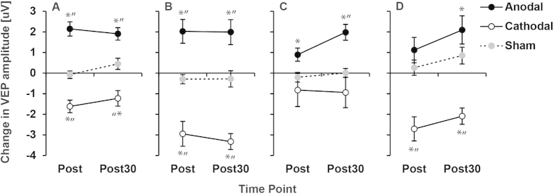
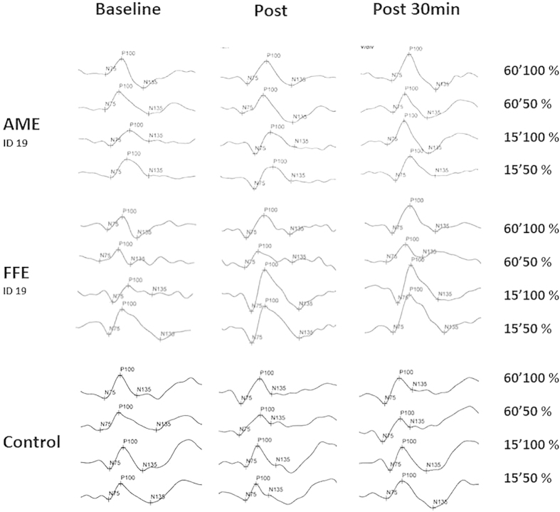
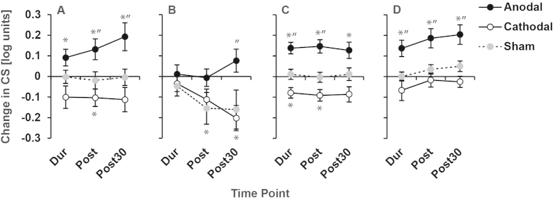
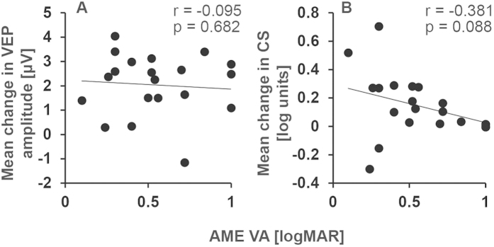
Amblyopia is a neurodevelopmental disorder of vision that occurs when the visual cortex receives decorrelated inputs from the two eyes during an early critical period of development. Amblyopic eyes are subject to suppression from the fellow eye, generate weaker visual evoked potentials (VEPs) than fellow eyes and have multiple visual deficits including impairments in visual acuity and contrast sensitivity. Primate models and human psychophysics indicate that stronger suppression is associated with greater deficits in amblyopic eye contrast sensitivity and visual acuity. We tested whether transcranial direct current stimulation (tDCS) of the visual cortex would modulate VEP amplitude and contrast sensitivity in adults with amblyopia. tDCS can transiently alter cortical excitability and may influence suppressive neural interactions. Twenty-one patients with amblyopia and twenty-seven controls completed separate sessions of anodal (a-), cathodal (c-) and sham (s-) visual cortex tDCS. A-tDCS transiently and significantly increased VEP amplitudes for amblyopic, fellow and control eyes and contrast sensitivity for amblyopic and control eyes. C-tDCS decreased VEP amplitude and contrast sensitivity and s-tDCS had no effect. These results suggest that tDCS can modulate visual cortex responses to information from adult amblyopic eyes and provide a foundation for future clinical studies of tDCS in adults with amblyopia.
Conflict of interest statement
The authors declare no competing financial interests.
Figures

References
-
- Daw N. Visual development. (Springer, 2014).
-
- Holmes J. M. & Clarke M. P. Amblyopia. Lancet 367, 1343–1351 (2006). - PubMed
-
- Hess R. F. & Howell E. R. The threshold contrast sensitivity function in strabismic amblyopia: evidence for a two type classification. Vision Res. 17, 1049–1055 (1977). - PubMed
-
- Ho C. S. & Giaschi D. E. Stereopsis-dependent deficits in maximum motion displacement in strabismic and anisometropic amblyopia. Vision Res. 47, 2778–2785 (2007). - PubMed
Publication types
MeSH terms
LinkOut - more resources
Full Text Sources
Other Literature Sources
Medical
