. 2016 Mar 1;27(5):838-47.
doi: 10.1091/mbc.E15-10-0707. Epub 2016 Jan 13.

A NIMA-related kinase, CNK4, regulates ciliary stability and length

Affiliations

A NIMA-related kinase, CNK4, regulates ciliary stability and length

Dan Meng et al. Mol Biol Cell. .

Abstract

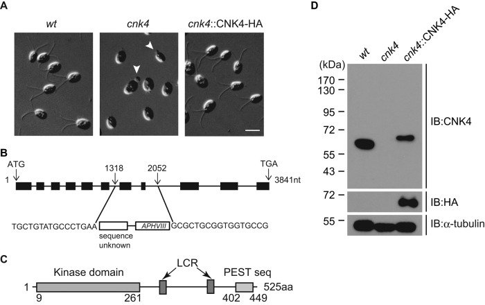
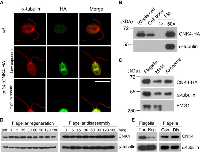
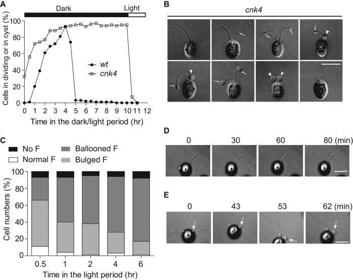
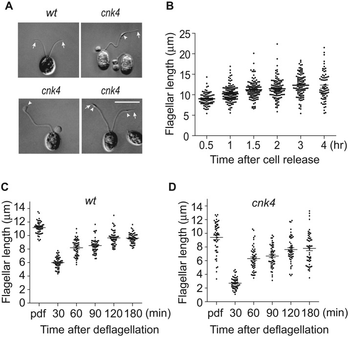
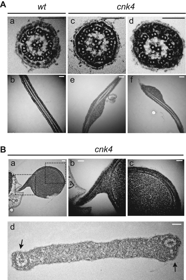
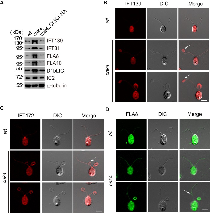
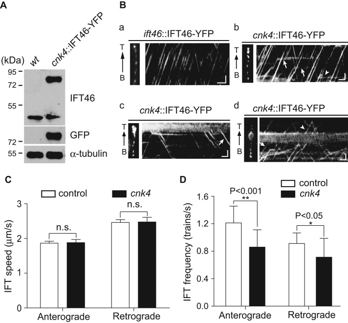
NIMA-related kinases (Nrks or Neks) have emerged as key regulators of ciliogenesis. In human, mutations in Nek1 and Nek8 cause cilia-related disorders. The ciliary functions of Nrks are mostly revealed by genetic studies; however, the underlying mechanisms are not well understood. Here we show that a Chlamydomonas Nrk, CNK4, regulates ciliary stability and length. CNK4 is localized to the basal body region and the flagella. The cnk4-null mutant exhibited long flagella, with formation of flagellar bulges. The flagella gradually became curled at the bulge formation site, leading to flagellar loss. Electron microscopy shows that the curled flagella involved curling and degeneration of axonemal microtubules. cnk4 mutation resulted in flagellar increases of IFT trains, as well as its accumulation at the flagellar bulges. IFT speeds were not affected, however, IFT trains frequently stalled, leading to reduced IFT frequencies. These data are consistent with a model in which CNK4 regulates microtubule dynamics and IFT to control flagellar stability and length.


Figures

FIGURE 1:
FIGURE 1:
cnk4 is defective in ciliogenesis. (A) Wild-type (wt), cnk4, and cnk4::CNK4-HA cells were imaged by DIC microscopy. Note that cnk4 cells are aflagellated or have flagellar buds (arrowheads). Bar, 10 μm. (B) Schematic illustration of the CNK4 gene, showing the replacement of exons 7 and 8 by foreign DNA fragment during random DNA insertion. The insertion site was identified by PCR and DNA sequencing. (C) The domain structure of CNK4. Numbers are amino acid positions. LCR, low-complexity region. (D) Western blot of whole-cell lysates from indicated strains with antibodies against CNK4, HA, and α-tubulin. No CNK4 was detected in the cnk4 mutant.
FIGURE 2:
FIGURE 2:
CNK4 is a flagellar protein. (A) CNK4 is localized to flagella and basal body. wt and cnk4::CNK4-HA cells were immunostained with anti-HA and anti–α-tubulin antibodies, respectively. High-exposure images show the flagellar location of CNK4. Bar, 10 μm. (B) cnk4::CNK4-HA cells were fractionated into cell body and flagella, followed by immunoblotting. Here 1×Fla (flagella) represents equal proportion of flagella to the cell body. (C) Isolated flagella of cnk4::CNK-HA cells were fractionated into membrane matrix (M+M) and axonemal fractions, followed by immunoblotting. FMG1 is a membrane protein. (D) wt cells were allowed to regenerate flagella after deflagellation (left) or treated with sodium pyrophosphate (NaPPi) to induce flagellar disassembly (right), followed by immunoblotting. (E) Flagella were isolated during flagellar regeneration after deflagellation or flagellar disassembly induced by NaPPi, followed by immunoblotting. Con, steady-state flagella; Dis, disassembling flagella; Reg, regenerating flagella.
FIGURE 3:
FIGURE 3:
cnk4 loses flagellar maintenance by forming flagellar bulges and balloons. (A) Cells were synchronized by using light/dark cycles under 5% CO2. cnk4 cells exhibited early onset of cell division, and the daughter cells were released upon lighting, whereas wt cells released daughter cells after completion of cell division. (B) DIC images of cnk4 cells show distinct flagellar phenotypes. The flagellar bulges and balloons are marked as arrows and arrowheads, respectively. Bar, 10 μm. (C) Quantification of different types of flagella after cells were released in the light period. F, flagella. (D, E) Movie stills showing flagellar bulge formation (D) and flagellar curling at the bulge formation site (E). Arrows mark the bulges. Note that the distal flagellum to the bulge is retracted into the bulge. Bars, 10 μm.
FIGURE 4:
FIGURE 4:
cnk4 loses flagellar length control. (A) DIC images of wt and cnk4 cells. cnk4 cells form extremely long flagella with bulge formation (arrowheads). Arrows mark the flagellar tips. Bar, 10 μm. (B) Flagellar length increase after cells are released from the mother cell wall of cnk4 cells. Cells were fixed at different times after cell release in the light period and imaged by DIC microscopy, followed by flagellar length measurement. Normal-looking flagella and flagella with bulges but not ballooned flagella were analyzed. (C, D) Flagellar regeneration of wt (C) and cnk4 (D) cells. Cells at 1 h in the light were deflagellated by pH shock to allow flagellar regeneration, followed by flagellar length measurement. Ballooned flagella were excluded from measurement. Note that cnk4 has a reduced rate of assembly before 30 min after deflagellation.
FIGURE 5:
FIGURE 5:
Electron microscopic analysis of cnk4 flagella. (A) Cross- and longitudinal sections of wt (a, b) and cnk4 (c–f) flagella. Note that cnk4 flagella accumulate amorphous materials between the membrane and the axoneme (d–f), whereas its substructures appear normal (d). Bars, 150 nm. (B) Flagellar balloon cross-sectioned to show curled axoneme (a–d) and degenerated axonemal microtubules (d, arrows). Dashed box images in (a) are enlarged and shown in (b) and (c), respectively. Bars, 150 nm.
FIGURE 6:
FIGURE 6:
Increase of IFT proteins in the cnk4 flagella. (A) Isolated flagella from wt, cnk4, and cnk4::CNK4-HA were analyzed by immunoblotting with antibodies against IFT proteins (IFT139, IFT-A; IFT81, IFT-B) and IFT motor subunits (FLA8, FLA10, and D1bLIC). IC2 and α-tubulin were used as controls. (B–D) wt and cnk4 cells were immunostained with antibodies against IFT139 (B), IFT172 (C), and FLA8 (D) antibodies. cnk4 flagella exhibit an overall increase of IFT proteins along the flagellar length compared with wt flagella. Flagellar bulges (arrows) are filled with IFT139, and IFT172 and FLA8 attach to the flagellar membrane. Bars, 10 μm.
FIGURE 7:
FIGURE 7:
cnk4 flagella exhibit normal IFT speed but reduced IFT frequency, with frequent IFT stalling. (A) Expression of yellow fluorescent protein (YFP)–tagged IFT46 in cnk4. Cells were analyzed by immunoblotting with antibodies against IFT46, GFP, and α-tubulin. (B) Kymographs of IFT46-YFP in the flagella of ift46::IFT46-YFP (a) and cnk4::IFT46-YFP (b–d) cells. Left, images of recorded flagella. (b) Kymograph from apparently normal flagellum. The arrows and arrowhead indicate stalled IFT in anterograde and retrograde directions, respectively. (c) Kymograph highlighting IFT turnaround at the bulged flagellar tip. Arrow, spontaneously initiated retrograde IFT. (d) Kymograph showing IFT at the flagellar bulge in the middle of the flagellum. Arrow, trapped IFT; arrowhead, IFT passing though the bulge. B, flagellar base; T, flagellar tip. Bars, 1 μm and 1 s. (C) The velocities of anterograde and retrograde IFT are not affected by cnk4 mutation. n.s., not significant (p > 0.05). (D) IFT frequencies of both directions in cnk4 flagella are reduced compared with those of wild-type flagella.

References

    1. Badano JL, Teslovich TM, Katsanis N. The centrosome in human genetic disease. Nat Rev Genet. 2005;6:194–205. - PubMed
    1. Berman SA, Wilson NF, Haas NA, Lefebvre PA. A novel MAP kinase regulates flagellar length in Chlamydomonas. Curr Biol. 2003;13:1145–1149. - PubMed
    1. Berthold P, Schmitt R, Mages W. An engineered Streptomyces hygroscopicus aph 7” gene mediates dominant resistance against hygromycin B in Chlamydomonas reinhardtii. Protist. 2002;153:401–412. - PubMed
    1. Bloodgood RA, Salomonsky NL. The transmembrane signaling pathway involved in directed movements of Chlamydomonas flagellar membrane glycoproteins involves the dephosphorylation of a 60-kD phosphoprotein that binds to the major flagellar membrane glycoprotein. J Cell Biol. 1994;127:803–811. - PMC - PubMed
    1. Bradley BA, Quarmby LM. A NIMA-related kinase, Cnk2p, regulates both flagellar length and cell size in Chlamydomonas. J Cell Sci. 2005;118:3317–3326. - PubMed

Publication types

MeSH terms

LinkOut - more resources