. 2016 Mar 1;27(5):744-56.
doi: 10.1091/mbc.E15-06-0373. Epub 2016 Jan 13.

Identification of DPY19L3 as the C-mannosyltransferase of R-spondin1 in human cells

Affiliations

Identification of DPY19L3 as the C-mannosyltransferase of R-spondin1 in human cells

Yuki Niwa et al. Mol Biol Cell. .

Abstract

R-spondin1 (Rspo1) is a secreted protein that enhances Wnt signaling, which has crucial functions in embryonic development and several cancers. C-mannosylation is a rare type of glycosylation and might regulate secretion, protein-protein interactions, and enzymatic activity. Although human Rspo1 contains 2 predicted C-mannosylation sites, C-mannosylation of Rspo1 has not been reported, nor have its functional effects on this protein. In this study, we demonstrate by mass spectrometry that Rspo1 is C-mannosylated at W(153) and W(156). Using Lec15.2 cells, which lack dolichol-phosphate-mannose synthesis activity, and mutant Rspo1-expressing cells that replace W(153) and W(156) by alanine residues, we observed that C-mannosylation of Rspo1 is required for its secretion. Further, the enhancement of canonical Wnt signaling by Rspo1 is regulated by C-mannosylation. Recently DPY19 was reported to be a C-mannosyltransferase in Caenorhabditis elegans, but no C-mannosyltransferases have been identified in any other organism. In gain- and loss-of-function experiments, human DPY19L3 selectively modified Rspo1 at W(156) but not W(153) based on mass spectrometry. Moreover, knockdown of DPY19L3 inhibited the secretion of Rspo1. In conclusion, we identified DPY19L3 as the C-mannosyltransferase of Rspo1 at W(156) and found that DPY19L3-mediated C-mannosylation of Rspo1 at W(156) is required for its secretion.


Figures

FIGURE 1:
FIGURE 1:
Establishment of an Rspo1-overexpressing cell line. (A) Schematic of human Rspo1. Rspo1 has two predicted C-mannosylation sites in the TSR1. The black box, two hatched bars, gray box, and two hexagonal shapes denote the signal peptide, furin repeats, TSR1, and putative C-mannosylation sites, respectively. (B) Amino acid sequence homology between human Rspo1-4 and mouse Rspo1. Two putative C-mannosylation sites are conserved in all Rspos. Conserved amino acids are indicated by asterisks, and putative C-mannosyltryptophan residues are underlined. (C) Rspo1 binds to cell surface HSPGs. Each cell line was cultured with or without 50 μg/ml soluble heparin, and all lysates and conditioned media were electrophoresed and immunoblotted with anti–c-myc and anti–α-tubulin. Rspo1 was secreted only after treatment with soluble heparin.
FIGURE 2:
FIGURE 2:
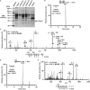
Identification of C-mannosylation sites in Rspo1. (A) Separation of recombinant Rspo1 from the conditioned medium of HT1080-Rspo1-MH cells. Secreted Rspo1-MH was purified with Ni-NTA agarose, and samples were electrophoresed on an SDS–polyacrylamide gel. The gel was visualized with CBB staining. (B–E) Identification of C-mannosylation sites in Rspo1. Samples were digested with trypsin and Asp-N, and the resulting peptides were analyzed by LC-MS/MS. The signal was observed in the chromatogram at m/z = 786.33 (B) and 705.31 (D). The ions at 9.97 min (m/z = 786.33) and 10.70 min (m/z =705.31) were further analyzed by LC-MS/MS, respectively (C, E). Indicated y ions were detected, and both W153 and W156 (C) or only W153 (E) of Rspo1 was C-mannosylated. C-mannosylation sites are underlined. *C, propionamide cysteine.
FIGURE 3:
FIGURE 3:
Effect of C-mannosylation on Rspo1 function. (A) Effect of C-mannosylation on Rspo1 secretion using CHO-K1 and Lec15.2 cells. CHO-K1 and Lec15.2 cells were transiently transfected with pCI-neo-Rspo1/N137Q-MH vector for 6 h and then cultured in serum-free medium with 50 μg/ml soluble heparin for 18 h. The protein samples were electrophoresed and immunoblotted with anti–c-myc and anti–α-tubulin. Signal intensities of Rspo1 were quantified and normalized to α-tubulin expression using ImageJ software. The Rspo1/α-tubulin ratio (CHO-K1) was defined as 1.0. (B, C) Establishment of mutant form of Rspo1 (W153 and W156 replaced by alanine residues; W153A/W156A: 2WA)–overexpressing HT1080 cell line, HT1080-Rspo1/2WA-MH. HT1080-neo (neo), HT1080-Rspo1-MH (wt), and HT1080-Rspo1/2WA-MH (2WA) cells were lysed, and aliquots of the cell lysates were electrophoresed and immunoblotted with anti–c-myc and anti–α-tubulin (B). Total RNA was isolated from each cell line, and semiquantitative (left) and quantitative (right) RT-PCR was performed (C). Equal amounts of exogenous Rspo1 in these cells were confirmed. ns, not significant. (D, E) Effect of C-mannosylation on intracellular trafficking. Cells were cultured with 50 μg/ml soluble heparin, stained with Hoechst 33258 (blue), anti–c-myc (green), and anti-KDEL (red; D) or anti-GRASP65 (red; E), and examined by fluorescence microscopy. Areas of overlapping stains are represented in yellow in the superimposed images. Bars, 10 μm. (F) Effect of C-mannosylation on Rspo1 secretion using a mutant Rspo1-overexpressing cell line. Cells were cultured with 50 μg/ml soluble heparin, and cell lysates and conditioned media were electrophoresed and immunoblotted with anti–c-myc and anti–α-tubulin. Signal intensities of Rspo1 were quantified and normalized to α-tubulin expression using ImageJ software. The Rspo1/α-tubulin ratio (wt) was defined as 1.0. (G) Effect of C-mannosylation on the kinetics of Rspo1 secretion. Cells were cultured with 50 μg/ml soluble heparin, and conditioned media were collected at the indicated times, electrophoresed, and immunoblotted with anti–c-myc (top). Protein bands were quantified by using ImageJ software (bottom). The secreted amount of wild-type Rspo1 at 24 h was defined as 100%. (H) Effect of C-mannosylation on Rspo1-mediated enhancement of Wnt signaling. Recombinant Rspo1 and Rspo1/2WA were purified from each cell line, and the amounts of proteins were equalized by Western blot (inset). 293T cells were transfected with TOPFlash or FOPFlash in the presence of 10% Wnt3a-conditioned medium and treated with equal amounts of purified Rspo1. After 24 h, luciferase activities were measured and normalized to Renilla luciferase. Non–C-mannosylated Rspo1 slightly enhanced Wnt signaling activity. Data shown are means ± SD. *p < 0.05 compared with FOPFlash of vehicle control treatment. **p < 0.05 compared with TOPFlash of vehicle control treatment. ***p < 0.05 compared with TOPFlash of vehicle control and wild-type Rspo1 treatments.
FIGURE 4:
FIGURE 4:
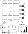
DPY19L3 is the C-mannosyltransferase of Rspo1 at W156. (A, B) Identification of C-mannosyltransferase of Rspo1. Human DPY19L1-L4– or empty vector (mock)–expressing Drosophila S2 cells were transiently transfected with pMT-Rspo1-MH, and protein expression was induced by 200 μM CuSO4 for 72 h. Rspo1-MH protein was purified by tandem affinity chromatography, heparin–Sepharose, and Ni-NTA agarose. The samples were digested with trypsin and Asp-N, and the resulting peptides were analyzed by LC-MS/MS. Monomannosylated peptide was observed only when the protein was produced in DPY19L3-expressing S2 cells (A). Unmannosylated and monomannosylated peptides derived from DPY19L3-expressing S2 cells were further analyzed by LC-MS/MS. Indicated y ions were detected, and signals resulting from the characteristic cross-ring cleavages were observed at the y7 and y8 ions in monomannosylated peptide (B). #W, C-mannosyltryptophan; *C, propionamide cysteine. (C) Effect of C-mannosylation of Rspo1 at W156 on Wnt signaling–enhancing activity. Recombinant Rspo1 proteins produced by mock- or DPY19L3-transfected S2 cells were purified, and the amounts of proteins were equalized by Western blot (inset). 293T cells were transfected with TOPFlash or FOPFlash in the presence of 10% Wnt3a-conditioned medium and treated with equal amounts of purified Rspo1 proteins. After 24 h, luciferase activities were measured and normalized to Renilla luciferase. C-mannosylated Rspo1 at W156 (produced by DPY19L3-expressing S2 cells) had increased activity compared with non–C-mannosylated Rspo1 (produced by mock-transfected S2 cells). Data shown are means ± SD. *p < 0.05 compared with FOPFlash of vehicle control treatment. **p < 0.05 compared with TOPFlash of vehicle control treatment. ***p < 0.05 compared with TOPFlash of vehicle control and mock-produced Rspo1 treatments.
FIGURE 5:
FIGURE 5:
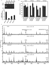
DPY19L3 is the C-mannosyltransferase of Rspo1 at W156 in human cells. (A) Expression of DPY19 members in HT1080-Rspo1-MH cells. Total RNA was isolated from HT1080-Rspo1-MH cells, and semiquantitative (top) and quantitative (bottom) RT-PCR were performed. Absolute copy number of mRNA transcript/10 ng of total RNA was calculated by quantitative RT-PCR. DPY19L2 expression was lower than the others. (B) Knockdown of DPY19L1, DPY19L3, and DPY19L4. Total RNA was isolated from each cell line, and quantitative RT-PCR was performed. Significant knockdown efficiency was observed for each siRNA against its target gene. (C) DPY19L3 is the C-mannosyltransferase of Rspo1 at W156 in human cells. HT1080-Rspo1-MH cells were treated with the indicated siRNAs, and conditioned media were collected. Recombinant Rspo1 was purified with Ni-NTA agarose, and the samples were digested with trypsin and Asp-N. The resulting peptides were analyzed by MALDI-TOF MS. siDPY19L3 changed the ratio of two peptides compared with siCtrl: the signal intensity from the dimannosylated peptide at W153 and W156 (m/z = 1919.0) declined, although that of the monomannosylated peptide at W153 (m/z = 1756.9) increased. #W, C-mannosyltryptophan; *C, propionamide cysteine. The ratio monomannosylated/dimannosylated Rspo1 was calculated from each peak area.
FIGURE 6:
FIGURE 6:
DPY19L3-mediated C-mannosylation of Rspo1 at W156 regulates its secretion. (A) Effect of knockdown of DPY19L1, DPY19L3, or DPY19L4 on Rspo1 secretion. HT1080-Rspo1-MH cells were treated with the indicated siRNAs and cultured in serum-free medium with 50 μg/ml soluble heparin, and cell lysates and conditioned media were electrophoresed and immunoblotted with anti–c-myc and anti–α-tubulin. Signal intensities of Rspo1 were quantified and normalized to α-tubulin expression using ImageJ software. The Rspo1/α-tubulin ratio (siCtrl) was defined as 1.0. (B, C) Effect of knockdown of DPY19L3 on Rspo1 secretion. HT1080-Rspo1-MH cells were treated with the indicated siRNAs. Total RNA was isolated from each cell line, and quantitative RT-PCR was performed (B). Each cell line was cultured in serum-free medium with 50 μg/ml soluble heparin, and cell lysates and conditioned media were electrophoresed and immunoblotted with anti–c-myc and anti–α-tubulin (C). Signal intensities of Rspo1 were quantified and normalized to α-tubulin expression using ImageJ software. The Rspo1/α-tubulin ratio (siCtrl) was defined as 1.0. (D) Effect of C-mannosylation of Rspo1 at W153 on Wnt signaling enhancing activity. HT1080-Rspo1-MH cells were treated with siGFP or siDPY19L3, and conditioned medium from each cell was collected. Rspo1 proteins were purified from the conditioned medium of siGFP- or siDPY19L3-treated cells, and the amounts of proteins were equalized by Western blot (inset). 293T cells were transfected with TOPFlash or FOPFlash in the presence of 10% Wnt3a-conditioned medium and treated with equal amounts of purified Rspo1 proteins. After 24 h, luciferase activities were measured and normalized to Renilla luciferase. W153-C-mannosylated Rspo1 (produced by siDPY19L3-treated cells) had almost same activity as wild-type Rspo1 (produced by siGFP-treated cells). Data shown are means ± SD. *p < 0.05 compared with FOPFlash of vehicle control treatment. **p < 0.05 compared with TOPFlash of vehicle control treatment. ns, not significant.

References

    1. Anastas JN, Moon RT. WNT signalling pathways as therapeutic targets in cancer. Nat Rev Cancer. 2013;13:11–26. - PubMed
    1. Buettner FF, Ashikov A, Tiemann B, Lehle L, Bakker H. C. elegans DPY-19 is a C-mannosyltransferase glycosylating thrombospondin repeats. Mol Cell. 2013;50:295–302. - PubMed
    1. Camp LA, Chauhan P, Farrar JD, Lehrman MA. Defective mannosylation of glycosylphosphatidylinositol in Lec35 Chinese hamster ovary cells. J Biol Chem. 1993;268:6721–6728. - PubMed
    1. Carlson CB, Lawler J, Mosher DF. Structures of thrombospondins. Cell Mol Life Sci. 2008;65:672–686. - PMC - PubMed
    1. Carmon KS, Gong X, Yi J, Thomas A, Liu Q. RSPO-LGR4 functions via IQGAP1 to potentiate Wnt signaling. Proc Natl Acad Sci USA. 2014;111:E1221–E1229. - PMC - PubMed

Publication types