. 2016 Mar 1;27(5):757-67.
doi: 10.1091/mbc.E15-10-0729. Epub 2016 Jan 13.

Inhibition of the FKBP family of peptidyl prolyl isomerases induces abortive translocation and degradation of the cellular prion protein

Affiliations

Inhibition of the FKBP family of peptidyl prolyl isomerases induces abortive translocation and degradation of the cellular prion protein

Pawel Stocki et al. Mol Biol Cell. .

Abstract

Prion diseases are fatal neurodegenerative disorders for which there is no effective treatment. Because the cellular prion protein (PrP(C)) is required for propagation of the infectious scrapie form of the protein, one therapeutic strategy is to reduce PrP(C) expression. Recently FK506, an inhibitor of the FKBP family of peptidyl prolyl isomerases, was shown to increase survival in animal models of prion disease, with proposed mechanisms including calcineurin inhibition, induction of autophagy, and reduced PrP(C) expression. We show that FK506 treatment results in a profound reduction in PrP(C) expression due to a defect in the translocation of PrP(C) into the endoplasmic reticulum with subsequent degradation by the proteasome. These phenotypes could be bypassed by replacing the PrP(C) signal sequence with that of prolactin or osteopontin. In mouse cells, depletion of ER luminal FKBP10 was almost as potent as FK506 in attenuating expression of PrP(C). However, this occurred at a later stage, after translocation of PrP(C) into the ER. Both FK506 treatment and FKBP10 depletion were effective in reducing PrP(Sc) propagation in cell models. These findings show the involvement of FKBP proteins at different stages of PrP(C) biogenesis and identify FKBP10 as a potential therapeutic target for the treatment of prion diseases.


Figures

FIGURE 1:
FIGURE 1:
Effect of FK506 treatment on PrPC expression. (A) N2a cells were treated with 20 μg/ml FK506 overnight, and the cell lysates were analyzed by immunoblotting for expression of endogenous PrP, BiP, and GAPDH. Here 0–2 indicate the different glycoforms of PrPC. (B) FK506 at the indicated concentrations was incubated overnight with N2a cells, and the expression of endogenous PrPC and MHC I was assessed by flow cytometry (n = 4, ±SD). (C) UPR activation was assessed by an XbpI mRNA splicing assay, using RNA isolated from N2a cells after overnight incubation with FK506. Tunicamycin (Tm) was used as a positive control, and DMSO served as a vehicle control. (D) HepG2 cells were incubated with 20 μg/ml CsA or 25 μg/ml FK506 overnight, followed by immunoblotting for PrPC, transferrin (Trf), albumin (Alb), PDI, BiP, and GAPDH. (E) N2a cells transiently transfected with a plasmid encoding hamster PrP were treated with DMSO or 20 μg/ml FK506 for 1 h. The cells were then starved in Met-free medium for 15 min and then radiolabeled with 150 μCi/ml [35S]Met for 20 min in the continued presence of the drug. PrPC was immunoisolated with mAb 3F4 and analyzed by SDS–PAGE and fluorography. Band doublets are due to the presence of both precursor and GPI-anchored forms of PrPC (Orsi et al., 2007).
FIGURE 2:
FIGURE 2:
FK506 treatment induces proteasome-mediated degradation of PrPC. (A) N2a cells were incubated with DMSO or 20 μg/ml FK506 for the indicated times, and PrPC expression was monitored by immunoblotting. (B) PrPC bands in A were quantified by densitometry, normalized to the GAPDH loading control, and expressed as a percentage of the protein present at the 0-min time point (n = 6, ±SEM). (C) N2a cells were digested with trypsin to remove surface PrPC and then treated with DMSO or 20 μg/ml FK506 for periods up to 22 h. The recovery of PrPC expression was monitored by immunoblotting. (D) Quantitation (n = 6–7, ±SEM). (E) The experiment in C was repeated with the addition of 5 μM bortezomib (bortez) to inhibit proteasome activity.
FIGURE 3:
FIGURE 3:
FK506 down-regulation of PrPC is not mediated by internalization and lysosomal degradation. (A) N2a cells were treated with 20 μg/ml FK506 or DMSO in combination with 25 μM chloroquine (Chloro) or 25 μM lactacystin (Lacta) overnight. PrPC expression was subsequently measured by immunoblotting. (B) N2a cells were incubated in serum-free DMEM with 20 μg/ml FK506 or DMSO in combination with 25 μM lactacystin and/or 0.025% trypsin (Tryp) overnight as indicated. PrPC expression was then measured by immunoblotting.
FIGURE 4:
FIGURE 4:
FK506 treatment enhances abortive translocation of PrPC. (A) N2a cells were transiently transfected with plasmids encoding hamster PrPC with either its wild-type (wt) signal sequence or that of osteopontin (Opn) or prolactin (Prl). After overnight treatment with DMSO or 20 μg/ml FK506 in the absence or presence of 5 μM bortezomib, cell lysates were immunoblotted with mAb 3F4 to selectively detect hamster PrPC. (B) N2a or HepG2 cells were treated overnight with DMSO, or 20 or 25 μg/ml FK506, respectively, in the absence or presence of 25 μM lactacystin. Cell lysates were split and digested with or without PNGase as indicated, followed by immunoblotting to detect PrPC. Arrows indicate the reduced mobility of PrPC that accumulates in FK506- and lactacystin-treated cells. (C) N2a cells were treated overnight with DMSO or 20 μg/ml FK506 in the absence or presence of 25 μM lactacystin as indicated. Cell lysates were split and subjected to PNGase digestion or left untreated. These samples were compared with N2a cells transiently transfected with control plasmid or plasmid encoding mouse ΔSP-PrPC, which lacks a signal sequence. The mobility of ΔSP-PrPC is indicated by the arrow. In all of these experiments, the SDS–PAGE gels were run for extended times to increase the resolution between the unglycosylated signal-cleaved and signal-uncleaved species. These forms of PrPC were typically not resolved under the SDS–PAGE conditions of the preceding figures.
FIGURE 5:
FIGURE 5:
FKBP10 depletion induces degradation of PrPC through proteasomal and lysosomal pathways. (A) Knockdowns (KDs) were performed over a period of 7 d in N2a cells using either FKBP10-specific siRNA or nontargeting control siRNA, and FKBP10 expression was assessed by immunoblot. Expression levels of PrPC, BiP, GAPDH, and CD90 were also monitored. For comparison, N2a cells were treated with 25 μg/ml FK506 overnight. (B) N2a cells were treated with FKBP10-specific siRNA or control siRNA for 3 d and subsequently treated with DMSO, 10 μM MG132 (MG), 25 μM chloroquine (Chl), or both (M+C) for 16 h. After deglycosylation with PNGase, FKBP10, PrPC and GAPDH levels were assessed by immunoblot. (C) Densitometric quantification of PrPC levels was carried out using ImageJ software (National Institutes of Health, Bethesda, MD). Values are expressed as a percentage of the level in control siRNA + vehicle–treated cells (n = 4 for FKBP10 siRNA + M+ C; n = 5 for all other conditions, ±SEM). *p < 0.005. (D) Knockdowns were performed over a period of 6 d in N2a cells using either FKBP10-specific siRNA or nontargeting control siRNA. In addition, the cells were transiently transfected on day 5 with plasmids encoding hamster PrPC with either its WT signal sequence or that of Prl or Opn. After deglycosylation with PNGase, the expression levels of FKBP10, PrPC, and GAPDH were monitored by immunoblot.
FIGURE 6:
FIGURE 6:
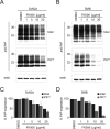
FK506 treatment effectively reduces PrPSc propagation. (A) ScN2a and (B) SMB cells were treated with DMSO or the indicated concentrations of FK506 for 6 d. The cells were lysed and split for the direct assessment of total PrP levels or for PK digestion and assessment of PK-resistant PrPSc levels. Total PrP and PrPSc were quantified and expressed as a percentage of the level in DMSO-treated control cells for (C) ScN2a and (D) SMB cell lines (n = 3, ±SD).
FIGURE 7:
FIGURE 7:
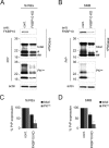
FKBP10 depletion effectively inhibits PrPSc propagation. FKBP10 knockdown was performed transiently using siRNA in (A) ScN2a and (B) SMB cells. The efficiencies of the knockdowns were validated in PNGase-treated lysates by immunoblotting for FKBP10 with actin as loading control. To evaluate PrP and PrPSc levels, cell lysates were split for the direct assessment of total PrP levels after PNGase treatment or for PK digestion and assessment of PK- resistant PrPSc. Here # and ## indicate C2 and C1 PrP fragments, respectively. Total PrP and PrPSc were quantified and expressed as a percentage of the level in control cells for (C) ScN2a and (D) SMB cell lines (n = 4, ±SD).

Similar articles

Cited by

References

    1. Barik S. Immunophilins: for the love of proteins. Cell Mol Life Sci. 2006;63:2889–2900. - PMC - PubMed
    1. Barnes AM, Cabral WA, Weis M, Makareeva E, Mertz EL, Leikin S, Eyre D, Trujillo C, Marini JC. Absence of FKBP10 in recessive type XI osteogenesis imperfecta leads to diminished collagen cross-linking and reduced collagen deposition in extracellular matrix. Hum Mutat. 2012;33:1589–1598. - PMC - PubMed
    1. Borchelt DR, Scott M, Taraboulos A, Stahl N, Prusiner SB. Scrapie and cellular prion proteins differ in their kinetics of synthesis and topology in cultured cells. J Cell Biol. 1990;110:743–752. - PMC - PubMed
    1. Brandner S, Isenmann S, Raeber A, Fischer M, Sailer A, Kobayashi Y, Marino S, Weissmann C, Aguzzi A. Normal host prion protein necessary for scrapie-induced neurotoxicity. Nature. 1996;379:339–343. - PubMed
    1. Bueler H, Aguzzi A, Sailer A, Greiner RA, Autenried P, Aguet M, Weissmann C. Mice devoid of PrP are resistant to scrapie. Cell. 1993;73:1339–1347. - PubMed

Publication types

MeSH terms

LinkOut - more resources