. 2016 Jan 13;19(1):55-66.
doi: 10.1016/j.chom.2015.12.004.

Structural Insights into Polymorphic ABO Glycan Binding by Helicobacter pylori

Affiliations

Structural Insights into Polymorphic ABO Glycan Binding by Helicobacter pylori

Kristof Moonens et al. Cell Host Microbe. .

Abstract

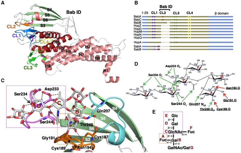
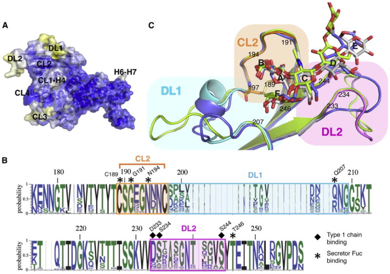
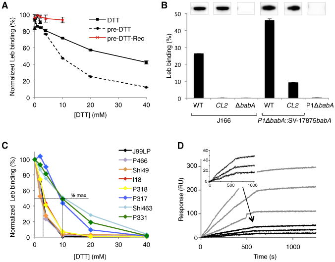
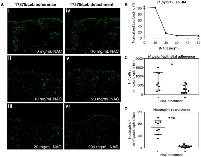
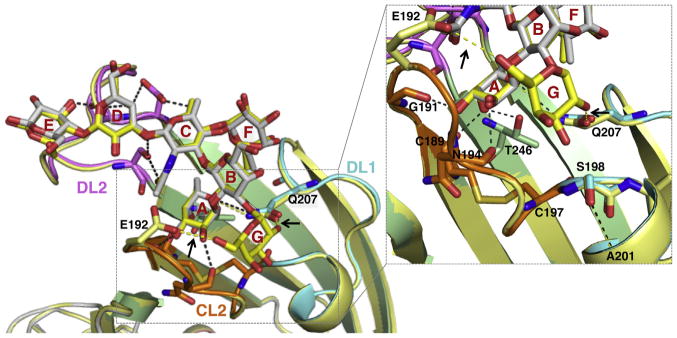
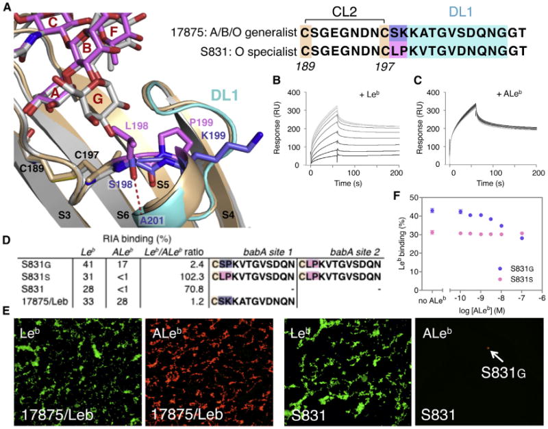
The Helicobacter pylori adhesin BabA binds mucosal ABO/Le(b) blood group (bg) carbohydrates. BabA facilitates bacterial attachment to gastric surfaces, increasing strain virulence and forming a recognized risk factor for peptic ulcers and gastric cancer. High sequence variation causes BabA functional diversity, but the underlying structural-molecular determinants are unknown. We generated X-ray structures of representative BabA isoforms that reveal a polymorphic, three-pronged Le(b) binding site. Two diversity loops, DL1 and DL2, provide adaptive control to binding affinity, notably ABO versus O bg preference. H. pylori strains can switch bg preference with single DL1 amino acid substitutions, and can coexpress functionally divergent BabA isoforms. The anchor point for receptor binding is the embrace of an ABO fucose residue by a disulfide-clasped loop, which is inactivated by reduction. Treatment with the redox-active pharmaceutic N-acetylcysteine lowers gastric mucosal neutrophil infiltration in H. pylori-infected Le(b)-expressing mice, providing perspectives on possible H. pylori eradication therapies.


Figures

Figure 1
Figure 1. BabAAD Interacts with Lewis b bg Antigens
(A) Schematic of the BabA architecture. Arrows indicate the aa 25–460 BabA adhesin domain fragment (BabAAD). Abbreviations: CL, cysteine-clasped loops; TM, predicted transmembrane domain; ID, Bab insertion domain (Figures S2 and S3). (B) ITC injection heats (upper) and normalized binding isotherm (lower) of the BabAAD titrated with Leb5. (C) SPR sensorgram of full-length BabA (solid and dashed lines show raw and fitted binding curves, respectively, for 500, 250, 125, 62.5, 31.3, and 15.7 nM BabA, from the top down; with a dissociation constant Kd = 3.9E-10 ± 0.9E-10 (M), an association rate constant ka = 6.1E5 ± 1.4E5 (M−1 s−1), and slow dissociation rate constant, kd = 2.3E-4 ± 0.8E-4 (s−1). (D) Similar SPR sensorgram of purified BabAAD binding to a Leb-coated chip; [BabA] as in (C). (E) Immunoblot detection of BabA from glutaraldehyde(GA) crosslinked H.pylori 17875/Leb bacterial cells; M, monomer, O, BabA oligomer. See also Figure S1.
Figure 2
Figure 2. Crystal Structure of BabAAD
(A) X-ray structure of strain 17875 BabAAD (for clarity, Nb-ER19 is not shown, see Figure S1D). Helices and strands are colored red and green, respectively; Cys-bound loops CL1 (Cys106-Cys135), CL2 (Cys189-Cys197), CL3 (Cys277-Cys299), and CL4 (Cys395-Cys423) are colored blue, orange, green, and yellow, respectively. Bab ID: residues 175–255 (Figures S2 and S3). (B) Schematic alignment of Cys-loop topology (vertical marks, colored as in Figures 1A and 1B) in the known or suspected Hop family adhesins. The α-helical ectodomain and β strand domains are colored brown and blue, respectively (sequence lengths not to scale, see Figure S2 for full MSA). (C) Structure of 17875 BabAAD (colored as in Figure 2) bound to Leb6 (Table S1). Leb6 and interacting amino acids (labeled) are shown in stick representation (O, N, and S atoms are colored red, blue, and yellow, respectively). Two binding subsites can be identified: the α1-2 fucose binding pocket (boxed orange) formed by CL2 and T246 in strand S6, and the type 1 chain binding region (boxed magenta) formed by the loop connecting strands S5 and S6 (i.e., DL2, see Figure 3). (D) H-bond network steering the 17875 BabAAD-Leb interaction. Side-chain- and main-chain-mediated H-bonds are depicted as green and red arrows, respectively. (E) Schematic of ABO Lewis b bg antigens (see Table S1), with monosaccharides labeled A–G.
Figure 3
Figure 3. Structure of BabAAD Bound to Lewis b bg H Hexasaccharide
(A) Solvent-accessible surface of BabAAD, with blue, white, and yellow corresponding to high, medium, and low sequence conservation in multiple sequence alignment of 237 publicly available BabA sequences. Four out of five regions of increased sequence diversity map to the same side of the adhesin: (i) the loop connecting CL1 and H4 (CL1-H4; residues 136–146); (ii and iii) two loops in the Bab ID, e.g., DL1 (Diversification Loop 1; residues 200–210, connecting CL2 and S4) and DL2 (residues 234–242, connecting S5 and S6) (Figures 2D and S3); and (iv) CL3 (residues 279–299). (B) Superimposition of strain 17875 BabAAD(colored as Figure 2C), with mutant BabAAD where the strain 17875 insertion domain is replaced by that of strain P437 (blue) or A730 (green). (C) Sequence conservation plot of the BabA insertion domain. Residues that interact with the secretor fucose and core 1 moiety are highlighted by asterisks and squares, respectively. See also Figure S3 and Table S2.
Figure 4
Figure 4. The CL2 Disulfide Bond Is Crucial for High-Affinity Leb Binding
(A) Normalized radioimmunoassay (RIA) of H. pylori strain 17875/Leb binding to 125I-labeled Leb in presence of increasing concentrations of DTT, added prior to (dotted lines) or coincubated with (solid lines) Leb. DTT-exposed bacteria resuspended in DTT-free buffer for 2 hr, show recovery of Leb binding (red line). Data points show mean ± SD, n = 2. (B) RIA Leb-binding of (left) the J166CL2 mutant (Cys189Ala and Cys197Ala), J166 WT, and J166ΔbabA; and (right) a babA deletion mutant of strain P1 (P1ΔbabA) and this strain conjugated with a shuttle vector expressing WT or CL2 mutant babA from strain 17875. Inlays show α-BabA immunoblots of the corresponding strains. Data points show mean ± SD, n = 2. (C) RIA experiment showing relative Leb binding of H. pylori clinical isolates preincubated with DTT. (D) SPR sensorgrams of the H. pylori strains with plasmid-based 17875 babA expression CL2 or WT BabA (black or gray response curves, respectively). Three dilutions of bacteria were flushed over immobilized Leb receptor conjugates, amplification of the CL2 curves are shown in inset. See also Figure S4.
Figure 5
Figure 5. Treatment of H. pylori cells with N-Acetylcysteine Blocks BabA Adherence
(A) FITC-labeled 17875/Leb bacteria binding to human gastric tissue sections. Prior to binding, bacteria were preincubated with 0, 10, or 20 mg/mL NAC (i, ii, and iii, respectively), resulting in 100%, 14%, and less than 5% adherent bacteria, respectively. In panels iv, v, and vi, tissue sections with bound bacteria are treated with 10, 20, or 200 mg/mL NAC, resulting in undetectable, 50%, and over 90% bacterial detachment, respectively. (B) Normalized RIA Leb-binding of H. pylori strain 17875/Leb when being subjected to increasing concentrations of N-acetylcysteine (0, 10, 20, 30, and 50 mg/mL) for 1 hr at 37°C. Data points show mean ± SD, n = 3. (C and D) H. pylori epithelial adherence and neutrophil recruitment in gastric epithelium of mice treated for 2 weeks with 0 (−) or 40 (+) mg/day NAC in their drinking water. Data points show, per animal, mean bacterial counts (n = 3) per mm of immunostained gastric epithelium (C; Figure S5D), or mean neutrophil counts (n = 3) per mm2 of immunohistostained gastric sections (D; Figure S5E). Statistical comparison of the groups produced a Welch-corrected t(9) = 2.29, *p = 0.0475; and t(7) = 7.559, ***p = 0.0001, respectively. Horizontal lines show sample mean ± SD, n = 8 (−NAC) and 9 (+NAC). See also Figure S5.
Figure 6
Figure 6. BabA Binding to bg A and B Glycans
Overlay of the structures of BabAAD bound to Leb6 (colored as Figure 3B) or BLeb7 (Table S1; yellow). Leb6 and BLeb7 are shown in stick representation, as are glycan-binding amino acids (shown for BabAAD Leb6 complex, and Glu192 and Gln207 for the BLeb7 complex). Hydrogen bonds present in both the Leb6 and BLeb7 interaction or specific to BabAAD:BLeb7 (see arrows) are shown in black or yellow dashed lines, respectively. Inset shows detail (rotated up by ∼45°) of the subpocket binding the bg B Gal. Complexes with ALeb5 or A6-1 are shown in Figure S6D.
Figure 7
Figure 7. Molecular Determinants of BabA bg Preference
(A) Structure of the BabA S831 DL1 grafted hybrid (BabAAD;DL1-S831; tan, positions 198–199 in magenta) in complex with Leb6 (magenta), and wild-type 17875 BabAAD bound to BLeb7 (light gray, with DL1 region in cyan and positions 198–199 in blue). In the specialist hybrid the replacement of Lys199 with Pro199 results in the inward rotation of residue 198. Replacement of Ser198 of 17875 by Leu198 causes a steric occlusion of the Gal or GalNAc determinants in BLeb or ALeb. (B and C) SPR sensorgrams of binding of BabAAD;DL1-S831 to a Leb-coated chip in the presence of competing soluble glycans: Leb5 (B) or ALeb5 (C), added in a 2-fold dilution series from 5 mM to 1 μM, colored gray to black. (D) RIA of Leb versus ALeb binding of a generalist (S831G[D]) and specialist clone (S831S), and the control strains S831 and 17875/Leb. Amino acid sequence ofthe DL1 region (shown starting at Cys197) of the babA allele in the two loci of the corresponding strains. Binding and sequencing data are representative for two independent S831G(D) and S831S clones isolated. (E) H. pylori 17875/Leb demonstrates the generalist phenotype and ability to bind both Alexa 488-labeled Leb (i) and Alexa 555-labeled ALeb (ii). Probing of theoriginal sweep population of the specialist H. pylori strain S831 by Alexa 555-labeled ALeb conjugate demonstrated by fluorescence microscopy the rarepresence of S831 bacterial cells of the generalist phenotype (F) Competition RIA where Leb binding to a S831G(D) (blue) and S831S (magenta) clone is performed in competition with nonradiolabeled ALeb. Increasing concentrations of ALeb titrate out 34.6% of the Leb binding signal, demonstrating the concomitant expression of a generalist and a specialist babA variant. Data points show mean ± SD, n = 3. See also Figure S7.

Comment in

  • H. pylori's BabA Embraces Change.
    Sweeney EG, Guillemin K. Sweeney EG, et al. Cell Host Microbe. 2016 Jan 13;19(1):5-7. doi: 10.1016/j.chom.2015.12.013. Cell Host Microbe. 2016. PMID: 26764590

Similar articles

Cited by

References

    1. Aird I, Bentall HH, Mehigan JA, Roberts JAF. The blood groups in relation to peptic ulceration and carcinoma of colon, rectum, breast, and bronchus; an association between the ABO groups and peptic ulceration. BMJ. 1954;2:315–321. - PMC - PubMed
    1. Alm RA, Bina J, Andrews BM, Doig P, Hancock RE, Trust TJ. Comparative genomics of Helicobacter pylori: analysis of the outer membrane protein families. Infect Immun. 2000;68:4155–4168. - PMC - PubMed
    1. Aspholm M, Kalia A, Ruhl S, Schedin S, Arnqvist A, Lindén S, Sjöström R, Gerhard M, Semino-Mora C, Dubois A, et al. Helicobacter pylori adhesion to carbohydrates. Methods Enzymol. 2006;417:293–339. - PMC - PubMed
    1. Aspholm-Hurtig M, Dailide G, Lahmann M, Kalia A, Ilver D, Roche N, Vikström S, Sjöström R, Lindén S, Bäckström A, et al. Functional adaptation of BabA, the H. pylori ABO blood group antigen binding adhesin. Science. 2004;305:519–522. - PubMed
    1. Boraston AB, Wang D, Burke RD. Blood group antigen recognition by a Streptococcus pneumoniae virulence factor. J Biol Chem. 2006;281:35263–35271. - PubMed

Publication types

MeSH terms